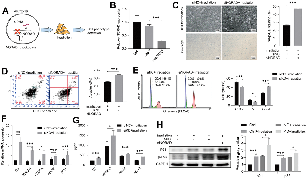
Figure 2.NORAD-knockdown aggravates irradiation-induced AMD markers of ARPE-19. ARPE-19 cell lines were transfected with siRNA of NORAD, after 24h, the cells were treated with irradiation, then observed for AMD phenotypes at different time periods. (A) Schematic diagram of experiment design to test the effect of NORAD on the irradiation-induced AMD model. (B) ARPE-19 were transfected with siNORAD or siNC. NORAD levels were analyzed through RT-qPCR. n = 3, ***P < 0.001. (C) ARPE-19 was observed cell morphology and stained to determine SA-β-gal. NORAD knockdown increased the number of positive-stained cells after they were treated with irradiation. Results were representative of three separate experiments. (100X, measure scar 50um) (D) NORAD-knockdown increased irradiation-induced cell apoptosis. The apoptosis rate was detected through flow cytometry by using annexin V-FITC/PI double staining. The apoptotic rate was analyzed in terms of the percentage of the lower and upper right quadrants. n = 3, ***P < 0.001. (E) NORAD silencing induced irradiation-treated cell cycle arrest at G2/M phase. Flow cytometry was used to detect the cell cycle distribution of irradiation-treated ARPE-19. n = 3, *P < 0.05, ***P < 0.001. (F) NORAD-knockdown increased mRNA expression of AMD markers in irradiation-treated ARPE-19 through qRT-PCR. n = 3, *P < 0.05, **P < 0.01, ***P < 0.001. (G) NORAD knockdown increased AMD markers of culture supernatant from irradiation-treated ARPE-19. n = 3, *P<0.05, ***P<0.001. (H) NORAD-knockdown increased the p-P53 and P21 expression observed through western blot. The ratio of p-P53 and P21 to GAPDH was analyzed with ImageJ. Values were shown as mean ± SD, n = 3, *P<0.05, ***P<0.001.