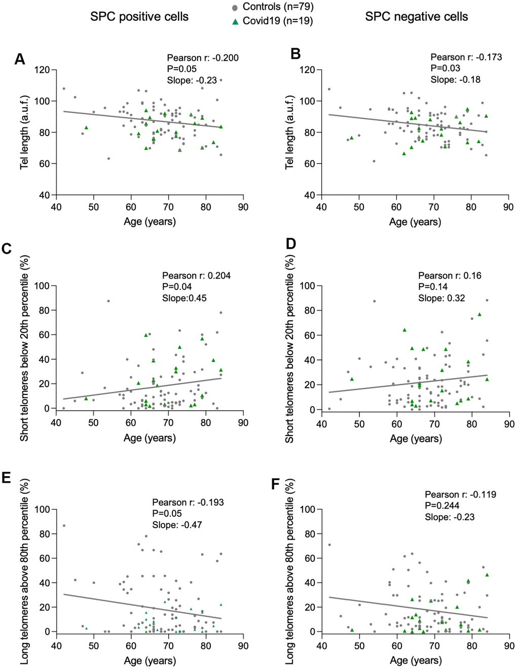
Figure 1.Progressive telomere shortening with age in lung cells. (A–F) Linear regression and Pearson correlation analyses between telomere intensity in pro-SPC positive (A) and pro-SPC negative cells (B), between percentage of short telomeres in pro-SPC positive (C) and pro-SPC negative cells (D), and between percentage of long telomeres in pro-SPC positive (E) and pro-SPC negative cells (F) and age in lung section from patients under study including control and COVID-19 samples. The Pearson r coefficient and the P value are indicated.