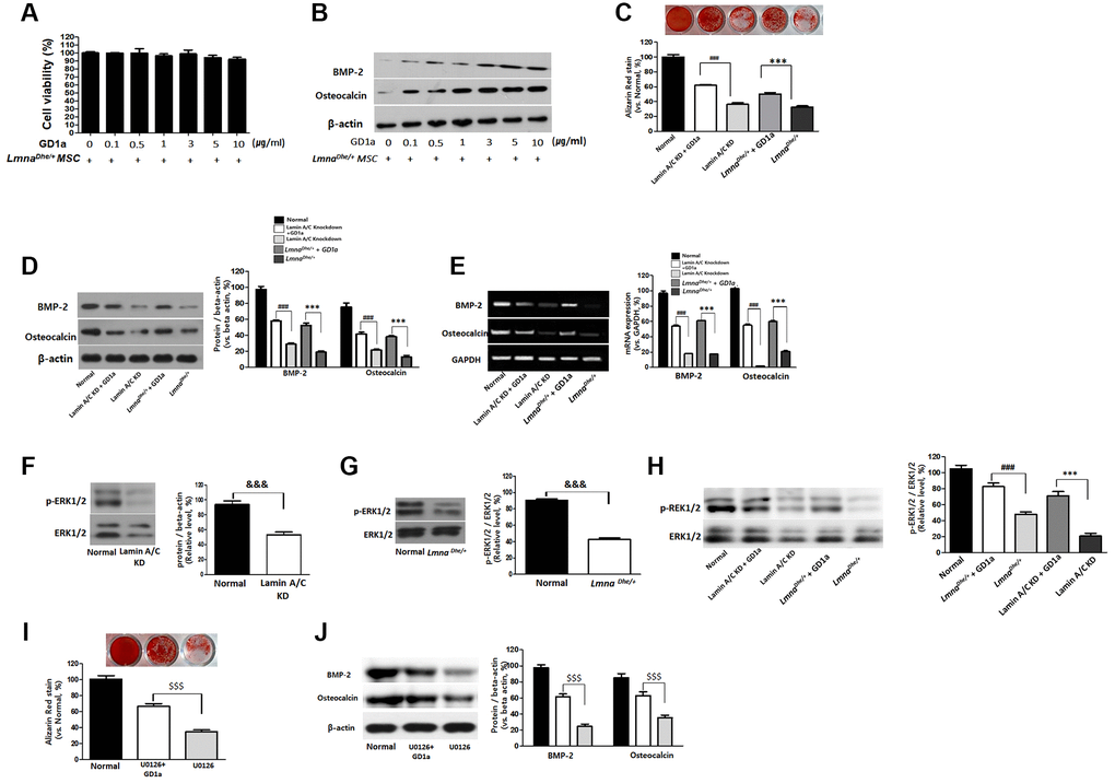
Figure 4.Increases of osteogenesis and ERK1/2 activation in GD1a was-treated Lmna dysfunction MSCs. Cell viability (A) and osteogenesis (B) in LmnaDhe/+ mutation MSCs were treated with GD1a. (C) Alizarin Red staining in GD1a was-treated Lmna dysfunction MSCs. (D) Increases of osteogenic proteins and (E) osteogenic genes in GD1a was-treated Lmna dysfunction MSCs compared with Lmna dysfunction MSCs. (F) Primary LmnaDhe/+ mutation MSCs were isolated from LmnaDhe/+ mutation mouse. (G) Lamin A/C was knocked down in mouse MSCs using siRNA. (H) Phosphorylation of ERK1/2 in Lmna dysfunction MSCs was treated with GD1a. (I) Alizarin Red staining in U0126-treated MSCs. (J) Increases of osteogenic proteins in GD1a-treated MSCs compared with pERK1/2-inhibited MSCs by U0126. Phosphorylation of ERK1/2 was determined by western blotting with anti-p-ERK1/2. ERK1/2 was used as a loading control. Values represent mean ± SD; &&&p < 0.001 indicates a significant difference from the normal MSCs; ***p < 0.001 indicates a significant difference from the LmnaDhe/+ mutant MSCs; ###p < 0.001 indicates a significant difference from the Lamin A/C KD MSCs. $$$p < 0.001 indicates a significant difference from the U0126-treated MSCs.