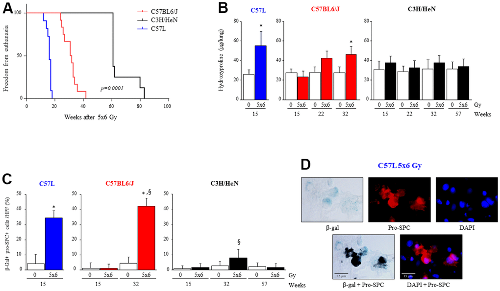
Figure 1.Varying susceptibility to radiation induced pulmonary fibrosis and pneumocyte senescence among three strains of mice. C57L, C57BL6/J, and C3H/HeN mice were exposed to 5 daily fractions of 6 Gy (5x6 Gy) of thoracic irradiation. At 15, 22, 32 and 57 weeks after irradiation, lung tissue was collected (n=5 mice per timepoint and condition). (A) Kaplan–Meier plot of freedom from euthanasia of irradiated mice (n≥10 mice per group) with comparison of curves using log rank test. (B) Hydroxyproline content was assessed in lung tissue at the indicated time point (in weeks) after irradiation. (C) Senescence associated-β-Galactosidase activity was assessed in lung samples collected at the indicated time points after irradiation, followed by immunocytochemical localization of pro-surfactant C. The percent of senescent AECII was scored. Columns: mean, error bars: +SD, *p<0.05 for comparison to 0 Gy for the corresponding strain and timepoint by ANOVA with Tukey’s correction. §p<0.05 for comparison to C57L lungs exposed to 5x6 Gy by ANOVA with Tukey’s correction. (D) Representative images of costaining of tissue sections for senescence associated-β-Galactosidase activity and pro-surfactant C in the C57L strain.