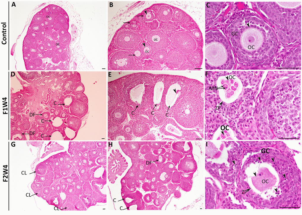
Figure 2.Histopathological changes in the ovaries from the first (F1W4) and the second generation (F2W4) compared to those in the control group (stained with H&E). (A–C) Photomicrographs at different magnifications of the same ovarian tissue section in females of control (CF1) showed normal structure containing normal growing follicles (arrows), normal oocytes (OC) (arrowheads), and a diminished number of pyknotic nuclei in granulosa cells (GC). (D, E) Photomicrographs of ovarian sections in treated females of F1W4 showing the presence of cysts (C), degenerated follicles (DF). (F) Segmented oocytes (arrowheads) enclosed by an irregular zona pellucida (ZP) with formation of micronucleus (MN). (G, H) Photomicrographs of ovarian sections in treated females of F2W4 showing a significant increase in growing follicle number and presence of corpora lutea (CL), there were a number of cysts (C) (arrows) and altered oocytes enclosed with abnormal zona pellucida (ZP), (I) in addition an elevation of pyknotic nuclei in granulosa cells (GC) (arrowheads). Scale bar = 60 μm.