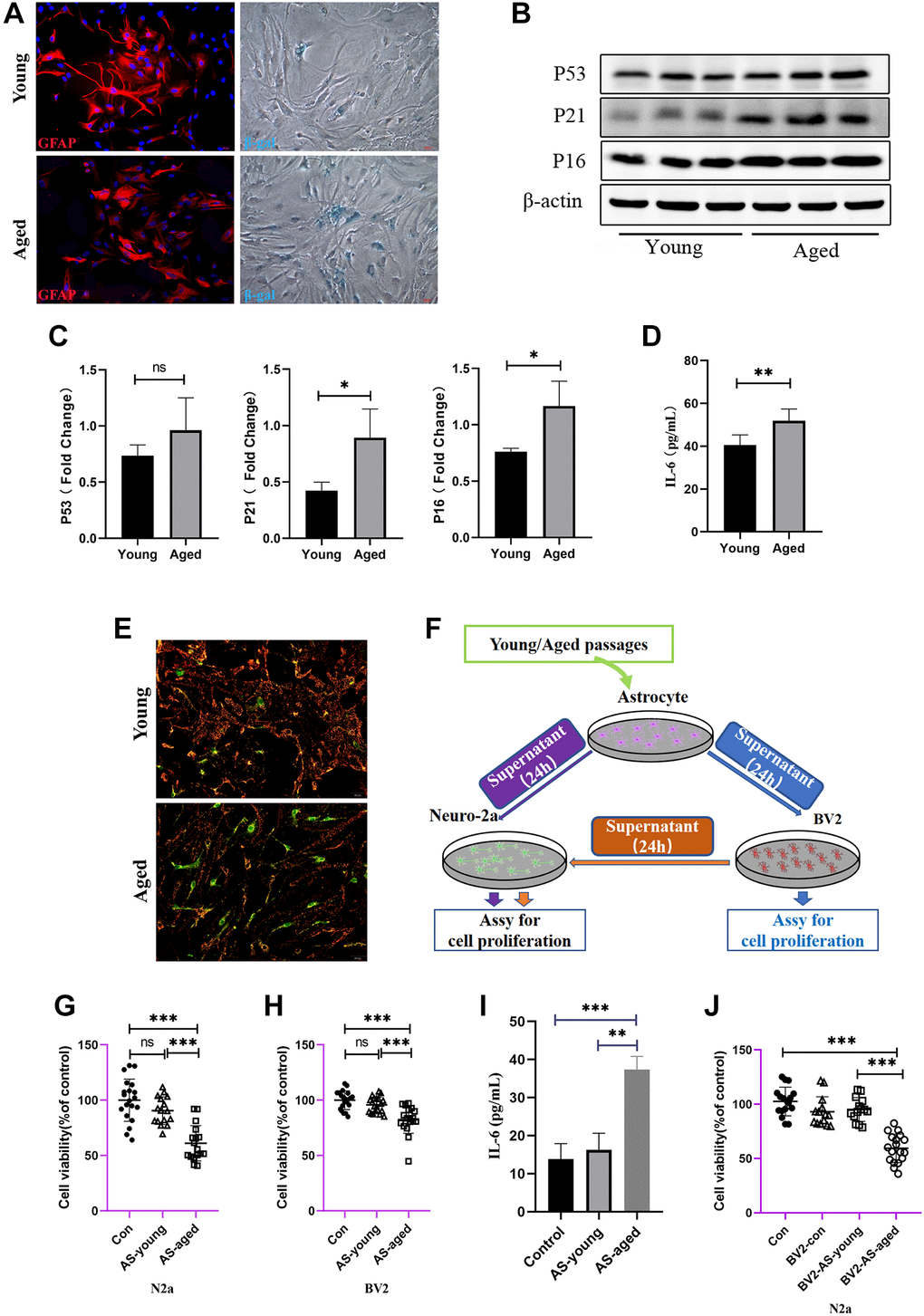
Figure 3.Astrocytes acquired senescent phenotype through serial passaging and SASP secretion may cause direct or indirect neuronal damage. (A) Immunofluorescent staining using GFAP antibody as astrocytes’ marker, and cell senescence staining of β-gal. (B) Representative immunoblot and (C) Quantitation of P53, P21, P16 and β-actin, in young and aged astrocytes. β-actin was a loading control and data are expressed relative to Young, n = 3. (D) Cytokine ELISA of IL-6 in culture medium released from Young and Aged astrocyte. (E) JC-1 staining. The red and green fluorescence reflects changes in the mitochondrial membrane potential of young and aged astrocytes, n = 3. (F) Scheme of conditioned media (CM) and cell viability assay by using young and aged astrocytes or astrocytes-CM treated BV2 cells. (G) Cell viability assay of N2a cells by treating with young and aged astrocytes’ supernatant. (H) Cell viability assay of BV2 cells by treating with young and aged astrocytes’ supernatant. (I) Cytokine ELISA of IL-6 in culture medium of BV2 treated with young/aged-astrocytes’ supernatant. (J) Cell viability assay of N2a cells by treating with supernatant of BV2 cells (treated with supernatant of young and aged astrocytes). All experiments were expressed as the mean +/−S.D, analyzed by ANOVA followed by Tukey’s test, *P < 0.05, **P < 0.01, ***P < 0.001, ns represents no significance.