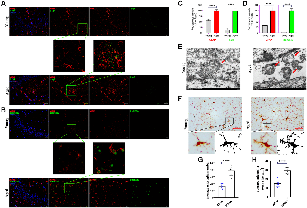
Figure 2.Senescent astrocytes occurred and accompanied with microglia activation. (A) Representative brain sections co-immunostained for GFAP (red) and β-gal (green) in young and aged mice (hippocampus). ×200 magnification. (B) Representative brain sections co-immunostained for GFAP (red) and P16INK4a (green) in young and aged mice (hippocampus). ×200 magnification. (C, D) Quantification of data from (A) and (B) (n = 6 mice/group). (E) Representative transmission electron microphotographs showing mitochondria in astrocytes from hippocampus of young and aged mice. Scale bar, 500 nm. (F) Brain sections immunostained for iba1 (activated markers of microglia). Scale bar, 50 μm. (G, H) Quantification of the average soma size and numbers of microglia in the CA3 hippocampal region. All experiments were expressed as the mean +/−S.D, analyzed by ANOVA followed by Tukey’s test, *P < 0.05, **P < 0.01, ***P < 0.001, ****P < 0.0001.