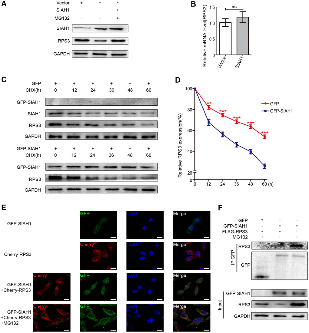
Figure 4.SIAH1 induces degradation of RPS3 protein. (A) A2780 cells were transfected with the Vector, SIAH1. Cells were treated with or without 10 μM MG132 for 6 h before cell lysis. and the resulting cell lysates were subjected to Western blotting. (B) RPS3 mRNA levels was detected in A2780 cells with SIAH1 overexpression. (C, D) The RPS3 protein half-life was assayed by using CHX (30 μg/ml) in HEK293T cells transfected with GFP or GFP-SIAH1 plasmid. The relative remaining RPS3 protein levels following CHX treatment at each time point were calculated accordingly. (E) Colocalization of RPS3 and SIAH1. The GFP-SIAH1 or Cherry-RPS3 plasmids were transfected into A2780 cells. Cells were treated with or without 10 μM MG132 for 6 h before cell lysis. GFP-SIAH1 was detected using a fluorescence microscope with an excitation wavelength of 488 nm. Cherry-RPS3 was detected with an excitation wavelength of 556 nm. The cell nuclei were stained with DAPI. Scale bar: 50 μm. (F) A2780 cells were transfected with plasmids as indicated, and the RPS3 protein was pull down with GFP antibody. Cell lysates were subjected to Western blotting. *p < 0.05, ***p < 0.001.