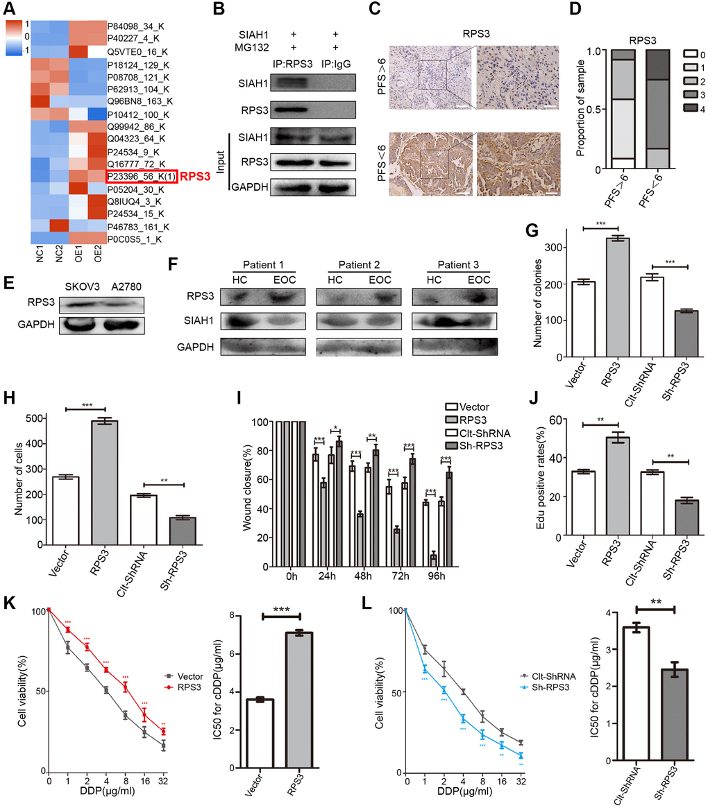
Figure 2.Identification of RPS3 as an interaction partner of SIAH1. (A) The heatmap showed that the protein level of RPS3 was down-regulated after SIAH1 overexpression in EOC cells. (B) Pull down of the RPS3 protein with SIAH1 antibody and IgG antibody in A2780 cells transfected with SIAH1. Cells were treated with 10 μM MG132 for 6 h before cell lysis. (C) Representative images of immunohistochemical staining for RPS3 in tumour specimens from ovarian cancer patients with PFS > 6 months vs. PFS < 6 months. Scale bar: 200 μm (left) and 100 μm (right). (D) The staining was assessed and scored on a scale of 0 (<5% staining) to 4 (>75% staining). The quantification of IHC staining (n = 24; PFS > 6, n = 12; PFS < 6, n = 12) was shown. (E) Protein level of RPS3 in SKOV3 and A2780 cells. (F) Protein levels of SIAH1 and RPS3 in serum samples from healthy controls (HC) and EOC patients (EOC). A2780 cells were separately transfected with Vector, RPS3, Clt-shRNA and sh-RPS3 for 48 h, the number of Cell Colonies was determined (G), the cell number from the Transwell assay was obtained (H), the Wound closure percentage was calculated (0, 24, 48, 72 and 96 h) (I), and the Edu positive rates (J), Cell Viability (K, L left panels), IC50 for cDDP (K, L right panels) were obtained. *p < 0.05, **p < 0.01, ***p < 0.001.