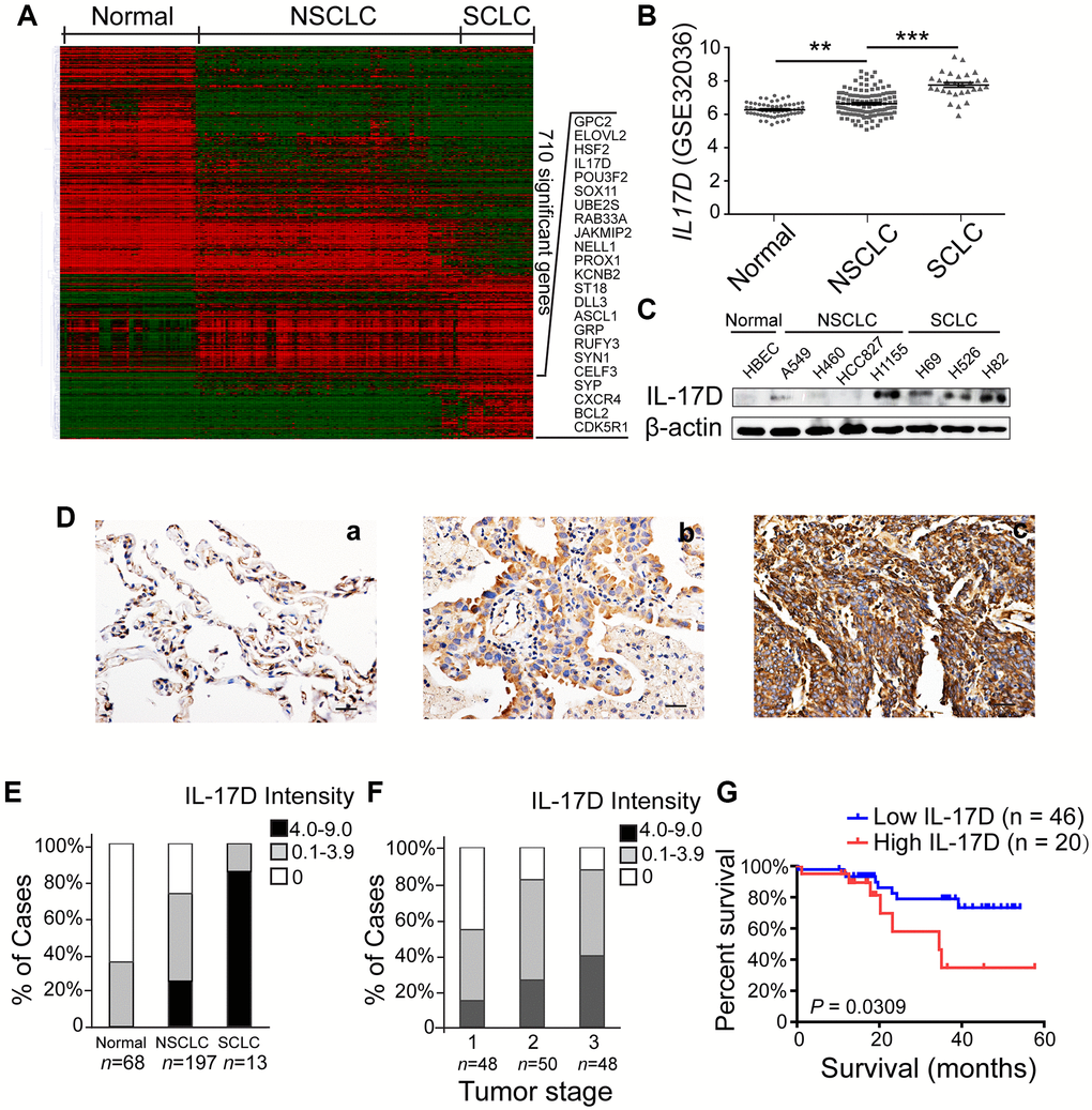
Figure 1.Interleukin (IL)-17D is highly expressed in lung cancer cell lines and tissues and correlates with poor prognosis in human lung cancer. (A) Heatmap of 856 differentially expressed transcripts (710 genes) among normal airway epithelial cell lines (Normal, n = 59), non-small cell lung cancer cell lines (NSCLC, n = 118), and small cell lung cancer cell lines (SCLC, n = 29). (B) RNA-seq data (GSE32036) of IL-17D gene expression of normal airway epithelial cells (n = 59) and NSCLC (n = 118) and SCLC (n = 29) cell lines (value = quantile-normalized and log2-transformed signal). **P < 0.01. ***P < 0.001. (C) Immunoblotting was performed to examine the expression level of IL-17D in normal, NSCLC, and SCLC cells. (D) Representative images of immunostaining with anti-IL-17D antibody in tissue sections of human lung tumor adjacent tissue (a), NSCLC tissue (b) and SCLC tissue (c). Scale bars are 20 μm. (E) The frequency of cases with no (0), low (0.1–3.9), or high (4.0–9.0) IL-17D staining stratified by immunohistochemically-defined lung cancer subtype. (F) The frequency of cases with no (0), low (0.1–3.9), or high (4.0–9.0) IL-17D staining stratified by tumor stage. (G) Kaplan-Meier survival rates for 66 subjects with lung cancer disease with low (staining scores < 2, n = 46, blue line) versus high (staining scores ≥ 2, n = 20, red line) IL-17D expression were compared. Median survivals were undefined months (low IL-17D) versus 34.52 months (high IL-17D; P = 0.0309).