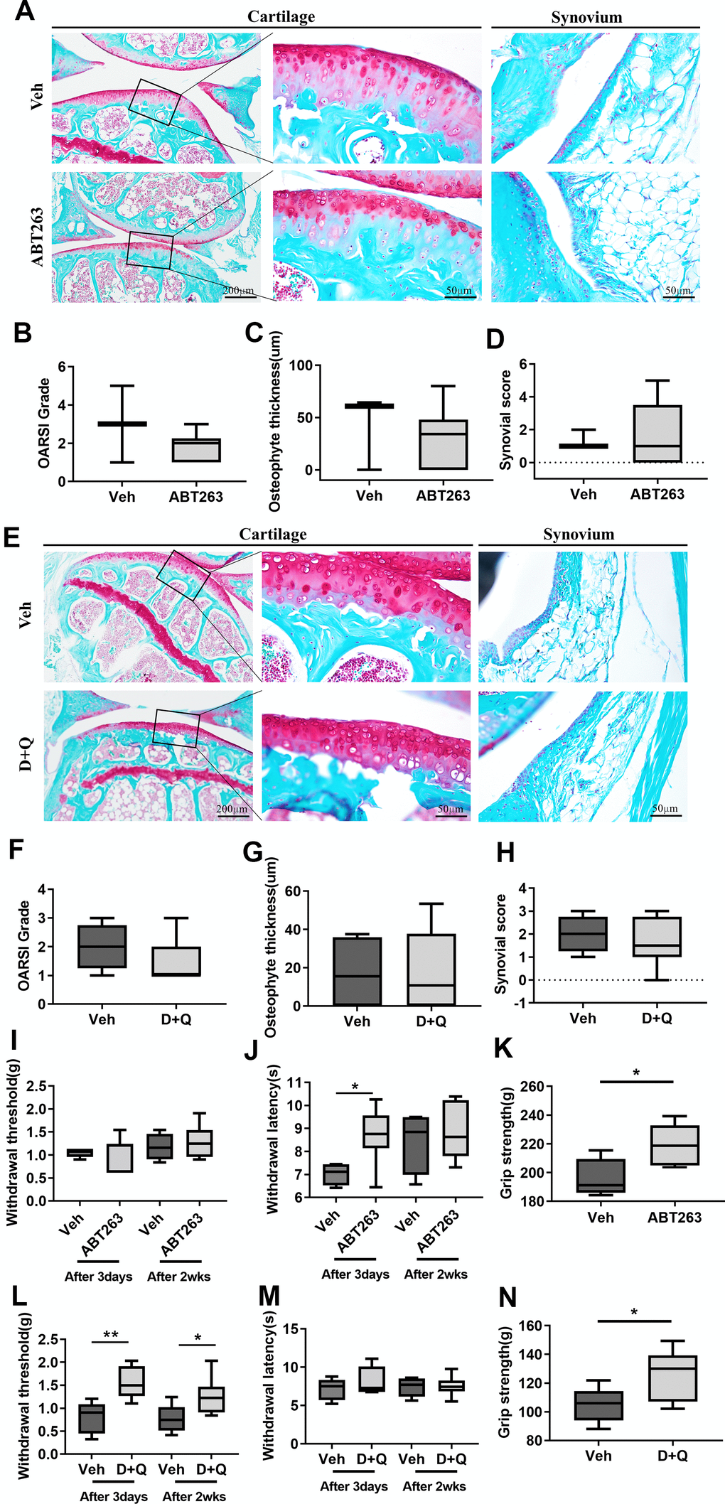
Figure 2.ABT263 and D+Q alleviate joint pain but do not affect cartilage degeneration during spontaneous OA progression. (A) Representative images of Safranin-O in the articular cartilage (left panel), higher magnification of the boxed regions (middle panel), and Safranin-O images of the synovium (right panel) of ABT263 and Veh-treated 21 to 22-month-old mice joints. (B) OARSI grade assessed by Safranin-O staining images in the medial tibial plateau, (C) osteophyte thickness of tibial articular cartilage, and (D) Krenn-synovitis score of ABT263 or Veh-treated aged mice (n = 3 mice for Veh, n = 6 mice for ABT263). (E) Representative images of Safranin-O staining in D+Q or Veh treated 21 to 22-month-old mice. Quantitative analysis of (F) OARSI grade, (G) osteophyte length, and (H) Krenn-synovitis score in the D+Q or Veh-treated aged mice (n = 4 for Veh, n = 8 for D+Q). (I) Von-Frey test to assess mechanical allodynia of the hind paws, (J) thermal hyperalgesia by Hargreaves apparatus, and (K) grip strength on day 3 and week 2 after the last dose of ABT263 treatment (n = 4 for Veh, n = 7 for ABT263). (L) Von-Frey test, (M) thermal hyperalgesia by Hargreaves apparatus, and (N) grip strength on day 3 and week 2 after the last dose of D+Q treatment (n = 5 for Veh, n = 8 for ABT263). Whisker plots represent the 10th and 90th percentiles, and the line corresponds to the median. * p < 0.05, ** p < 0.01; Unpaired Student’s t-test. Scale bars are shown in each image.