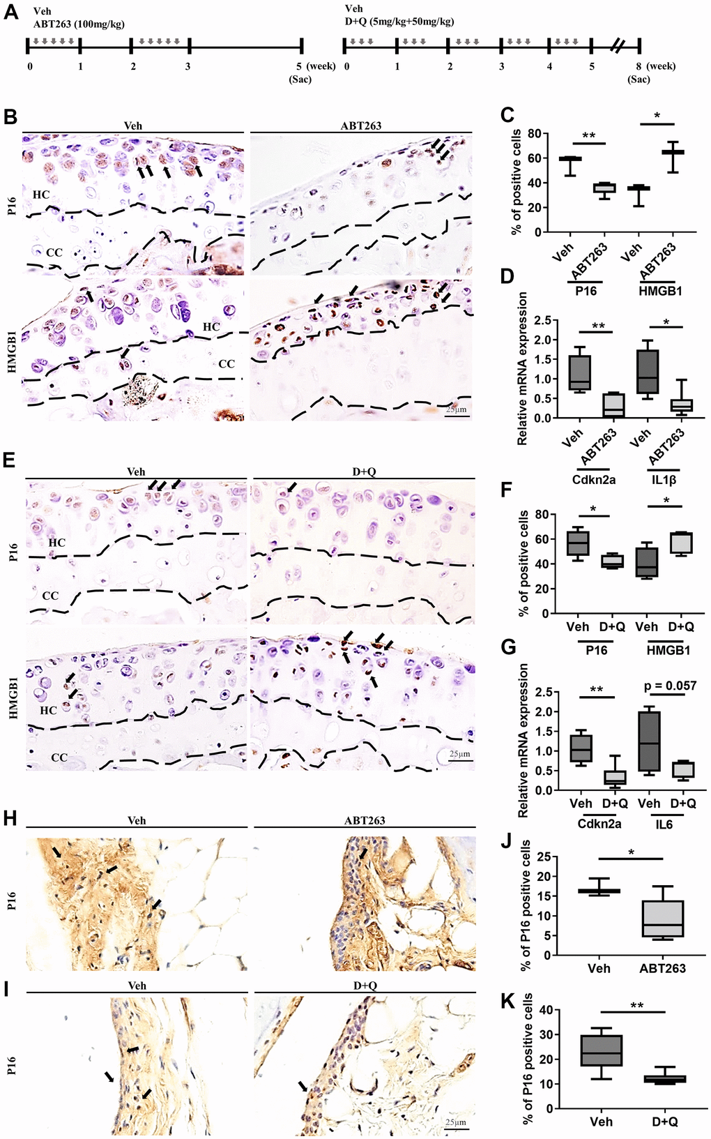
Figure 1.ABT263 and D+Q deplete senescent cells in articular joints of aged mice. (A) Timeline of the ABT263 and Dasatinib (D) + Quercetin (Q) treated aged mouse model of spontaneous OA. We orally administered ABT263 at 100 mg/kg or Vehicle (Veh) to 21 to 22-month-old mice five times biweekly for 4 weeks. The 21 to 22-month-old mice were given oral D + Q (5 mg/kg Dasatinib plus 50 mg/kg Quercetin) or Veh three times every week for 5 weeks. (B) Representative immunostaining images of p16 and HMGB1 + cells (arrows) in the articular cartilage of the ABT263 or Veh-treated aged mice. HC, hyaline cartilage; CC, calcified cartilage. (C) Quantification of p16 + SnCs and non-SnCs positive for nuclear HMGB1 in the articular cartilage (n = 3 mice for Veh, n = 5 mice for ABT263). (D) mRNA expression levels of Cdkn2a and IL1β are validated by qRT-PCR analysis in the aged mice administered ABT263 or Veh (n = 4 for Veh, n = 7 for ABT263). (E) Representative immunostaining for p16 and HMGB1 positive cells (arrows) in the articular cartilage of D+Q or Veh treated aged mice. (F) Quantification of p16 and HMGB1 + cells in the articular cartilage (n = 5 per group). (G) Quantification of mRNA levels for Cdkn2a and IL6 by qRT-PCR in the joints of D+Q or Veh-treated aged mice (n = 4 for Veh, n = 7 for D+Q). Representative images of immunostaining for p16 in the synovium of (H) ABT263 or Veh-treated or (I) D+Q or Veh-treated in the articular knee joint of the aged mice. Percentage of p16+ cells in the synovial membrane of (J) ABT263 or Veh-treated (n = 3 for Veh, n = 6 for ABT263) or (K) D+Q or Veh-treated mice (n = 5 for Veh, n = 6 for D+Q). Whisker plots represent the 10th and 90th percentiles, and the line corresponds to the median. * p < 0.05, ** p < 0.01; a two-tailed Student’s t-test (unpaired). Scale bars, 25 μm.