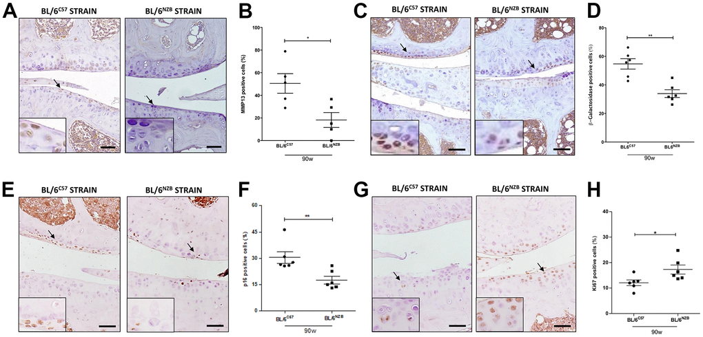
Figure 3.Age-associated changes in cartilage expression of senescence and proliferation markers in BL/6C57 and conplastic (BL/6NZB) mice. (A) Representative images of medial compartment of knee joints from BL/6C57 and conplastic (BL/6NZB) mice at 90 weeks of age stained with matrix metalloproteinase 13 (MMP13). (B) Quantitative analysis of MMP13-positive cells of knee joints from BL/6C57 and conplastic (BL/6NZB) mice at 90 weeks of age. (C) Representative images of medial compartment of knee joints from BL/6C57 and conplastic (BL/6NZB) mice at 90 weeks of age stained with β-Galactosidase. (D) Quantitative analysis of β-Galactosidase-positive cells of knee joints from BL/6C57 and conplastic (BL/6NZB) mice at 90 weeks of age. (E) Representative images of medial compartment of knee joints from BL/6C57 and conplastic (BL/6NZB) mice at 90 weeks of age stained with p16. (F) Quantitative analysis of p16-positive cells of knee joints from BL/6C57 and conplastic (BL/6NZB) mice at 90 weeks of age. (G) Representative images of medial compartment of knee joints from BL/6C57 and conplastic (BL/6NZB) mice at 90 weeks of age stained with Ki67. (H) Quantitative analysis of Ki67-positive cells of knee joints from BL/6C57 and conplastic (BL/6NZB) mice at 90 weeks of age. Original magnification: 20×. Scale bar, 50 μm. Black arrow indicates positively stained chondrocyte. Chondrocyte magnification (40×) is shown in the bottom-left corner of the images. Graphs represent means ± SEM; n=5 in BL/6C57 and n=5 in conplastic (BL/6NZB) mice at 90 weeks of age for MMP13, and n=6 in BL/6C57 and n=6 in conplastic (BL/6NZB) mice at 90 weeks of age for β-Galactosidase, p16, and Ki67. *p<0.05, * *p<0.01 by non-parametric Mann-Whitney test.