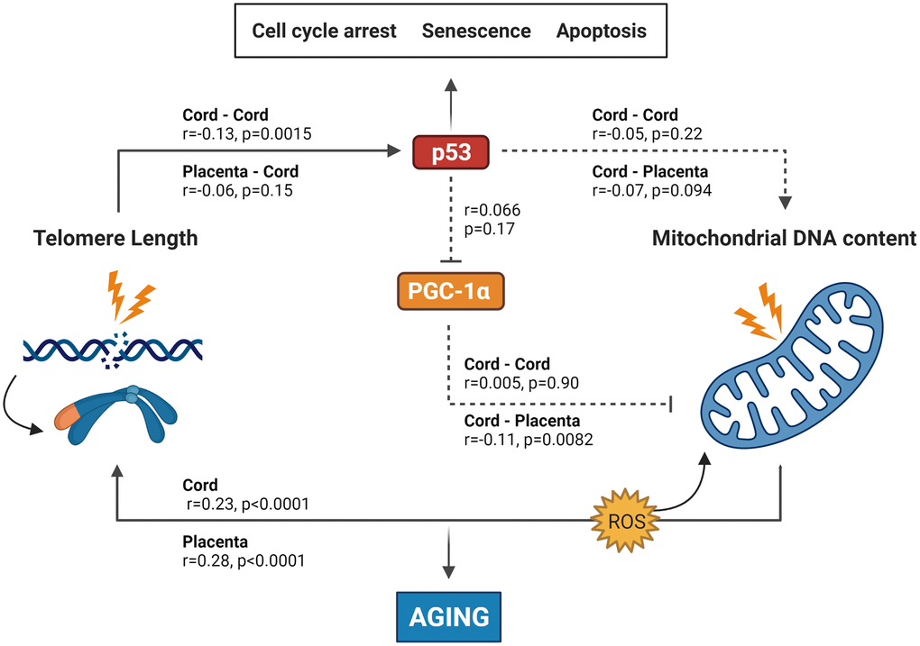
Figure 1.Summary of the results found in this study integrated into the experimentally based telomere-mitochondrial axis of biological aging hypothesis. DNA damage and telomere shortening activate p53 leading to growth arrest, senescence or apoptosis. p53 might also impair mitochondrial function and mitochondrial DNA content indirectly through suppression of PGC-1α – one of the master regulators of the mitochondria – leading to mitochondrial comprise and increased ROS levels, which leads to more DNA damage including telomere shortening. p53 and PGC-1α could therefore be central players in the association between telomere length and mitochondrial DNA content and subsequently in the aging process. Solid lines represent significant associations between age-related or protein markers, while non-significant associations are represented by dotted lines. p53 and PGC-1α levels were only measured in cord blood, while TL and mtDNAc were measured in both cord blood and placental tissue. Abbreviations: p53: tumor suppressor protein 53; PGC-1α: peroxisome proliferator-activated receptor gamma co-activator 1 alpha protein. Figure based on the experimental work of Sahin et al. [5, 33].