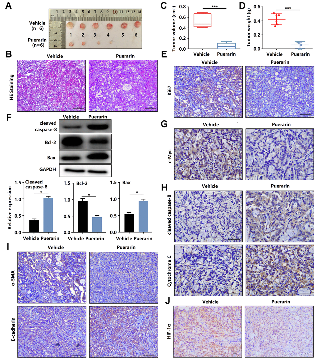
Figure 3.Puerarin inhibits the tumor growth and metastasis of PDAC in the animal xenograft model. (A) Effects of puerarin on morphologic changes in the experimental groups. (B) Pathological results of H&E staining for PDAC in tissues of the model group. Bar = 50 μm. (C) Effect of puerarin on the volume of tumors in the animal xenograft model. (D) Effects of puerarin on tumor weight. (E) IHC staining for Ki67 in the puerarin-treated model. Bar = 50 μm. (F) IHC staining for c-Myc in the puerarin-treated model. Bar = 50 μm. (G) Protein expression of Cleaved caspase-8, Bax and Bcl-2 in PDACs in different groups. (H) IHC staining for Cleaved caspase-8 and cytochrome C in the puerarin-treated model. Bar = 50 μm. (I) IHC staining for E-cadherin and α-SMA in the puerarin-treated model. Bar = 50 μm. (J) IHC staining for HIF-1α in the puerarin-treated model. Bar = 50 μm. The data are presented as the mean ± standard deviation, and were analyzed by a two-sided Student’s t-test. *p < 0.05 and ***p < 0.001.