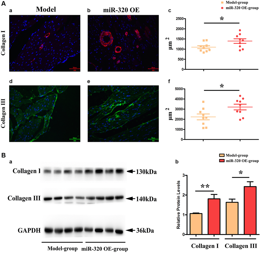
Figure 4.MiR-320 OE increased the type I and III collagen synthesis; (Aa–d) Representative pictures of type I and type III collagen staining in the left ventricular tissues; (Ac–f) The expression levels of type I and type III collagen synthesis; P < 0.05; miR-320 OE vs. model; (Ba) Representative Western blots in the left ventricular tissues to show the collagenous fibers synthesis; (Bb) The quantitative analysis of type I and type III collagen; P < 0.05; miR-320 OE vs. model.