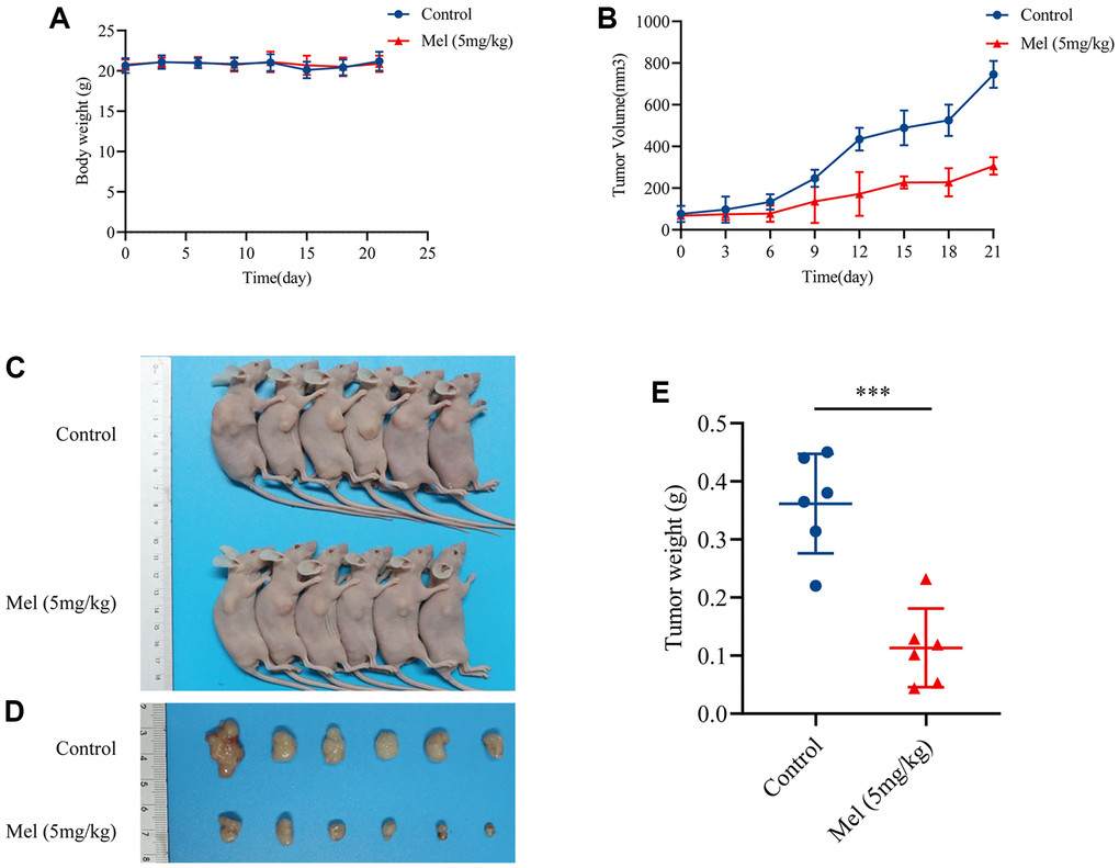
Figure 6.Melatonin inhibited GBC-SD cells proliferation in vivo. (A) Body weights of all mice were recorded every three days (n = 6). (B) The tumor volume was measured every three days (n = 6). (C, D) The pictures of mice and harvested tumors (n = 6). (E) Tumor weight measurements (n = 6). Data are presented as mean ± SD. Mel, melatonin; ***P < 0.001.