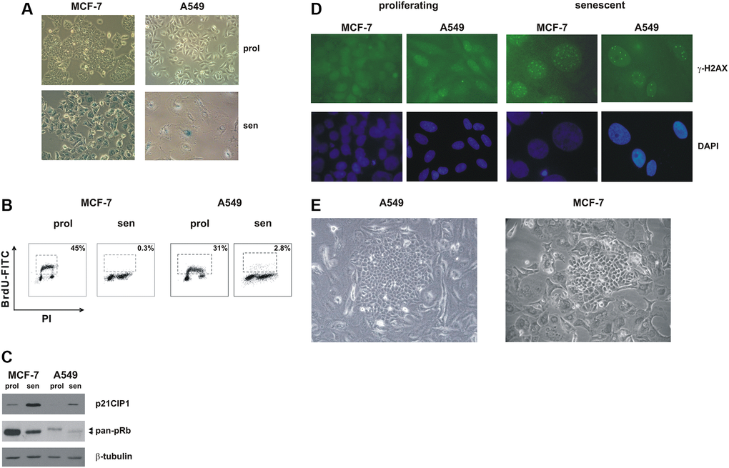
Figure 1.Premature senescence in MCF-7 and A549 cells. MCF-7 and A549 cells were treated with doxorubicin for 72 h. Cells were extensively washed and analyzed 7 days after release from the drug. (A) Morphological alterations and SA-β-gal staining in drug-induced senescent cells. Proliferating cells and doxorubicin-induced senescent cells were stained to detect SA-β-gal activity. Phase contrast microscopy images were captured using Canon powershot G6 camera at 10x magnification, 6× digital zoom. (B) Representative flow cytometric data. Proliferating and senescent MCF-7 and A549 cells were incubated with 5-bromo-2-deoxyuridine (BrdU), for 30 min and 1 hour, respectively. The number of BrdU-labelled cells was determined and the percentage is shown in the chart. (C) Accumulation of p21CIP1 and hypophosphorylated pRb protein in drug-induced senescent cells. Filters were stripped and reprobed with β-tubulin antibodies as a loading control. (D) Proliferating and doxorubicin-induced senescent MCF-7 and A549 cells were immunostained with an anti-γ-H2AX monoclonal antibody followed by secondary fluorescein-conjugate antibodies. Nuclei were stained with DAPI. Samples were visualized on a Zeiss Axioplan fluorescent microscope at 63x magnification. (E) Representative phase contrast microscopy images of clones that evade senescence; note the flat and enlarged morphology of TIS cells surrounding the escaped clone. Phase contrast microscopy images were captured using Canon powershot G6 camera at 20× magnification, 6× digital zoom.