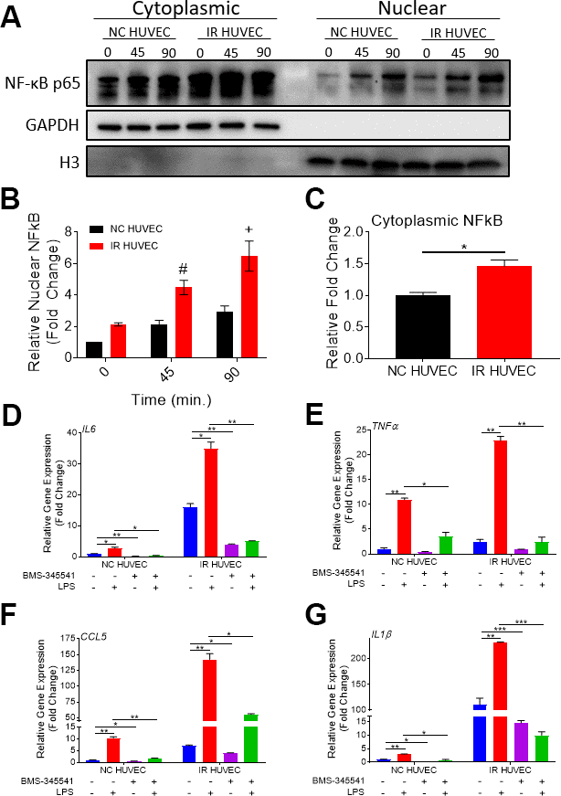
Figure 6.Regulation of senescence-associated hyper-activation via NF-κB pathway. (A–C) IR HUVEC exhibit higher baseline expression and activation of NF-κB when compared to NC HUVEC. Representative western-blot images (A) (the middle line is the molecular weight markers), densitometry based quantitative analysis of nuclear fraction (B) and cytoplasmic fraction (C) of NF-κB p65 in NC HUVEC and IR HUVEC stimulated with LPS (30 ng/ml) for 0-90 min (n = 3; mean ± SEM; * p<0.05, # p <0.01, + p<0.001 vs. NC HUVEC). Histone H3 and GAPDH were used as the loading control for nuclear and cytoplasmic proteins, respectively. (D–G) NF-κB inhibition attenuates the expression of IL6 (D), TNFα (E), CCL5 (F), and IL1β (G) mRNA in IR HUVEC. NC HUVEC and IR HUVEC were treated with LPS (30 ng/ml) or the NF-κB inhibitor BMS-345541 (10 μM) or their combination for three hours followed by mRNA analysis. Gene expression in unstimulated NC HUVEC was used as baseline and GAPDH was used as endogenous control. (n = 3; mean ± SEM; * p<0.05, ** p<0.01, *** p<0.001).