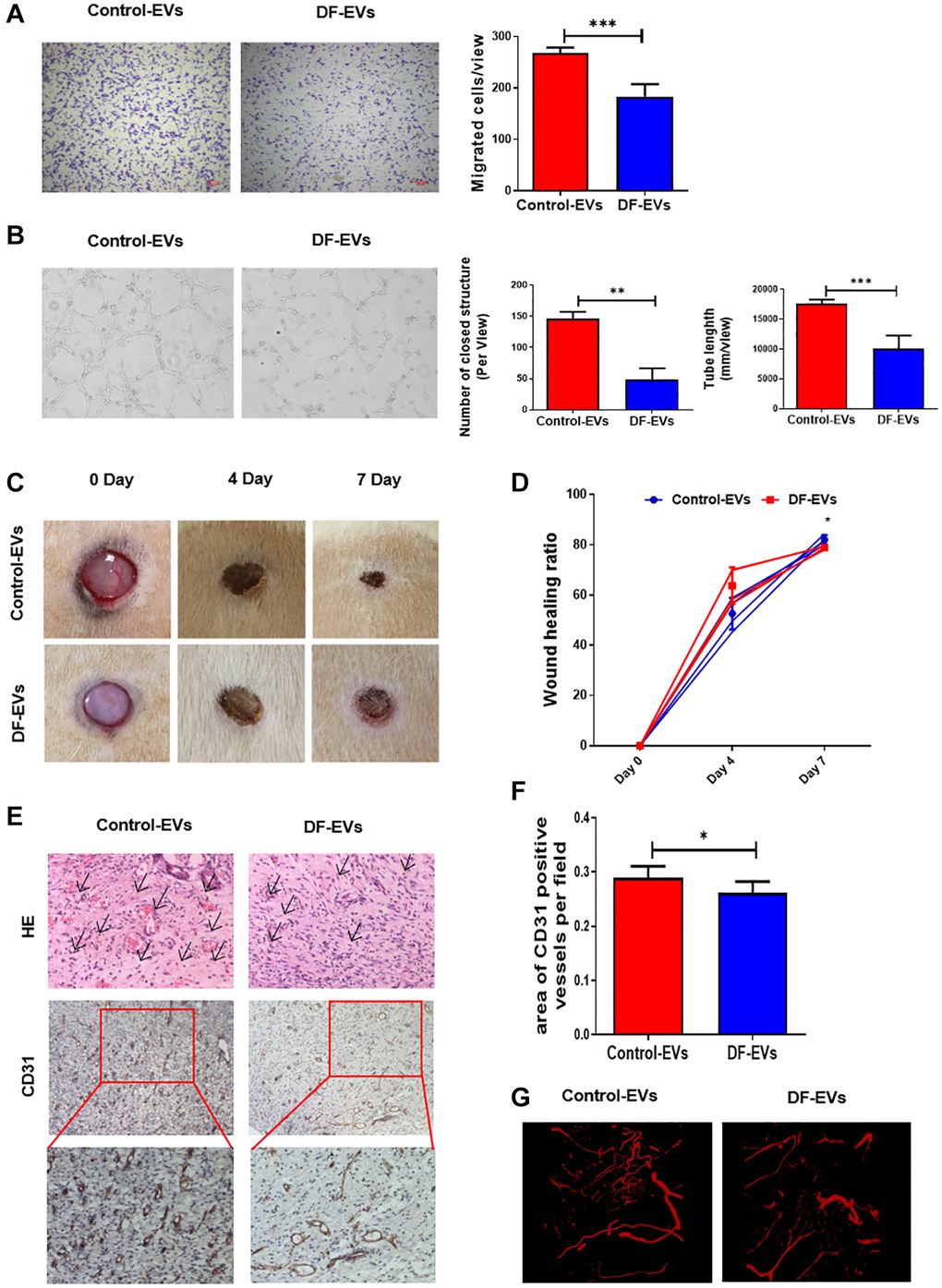
Figure 2.DF-EVs inhibited HUVEC angiogenesis in vitro and in vivo. (A) Transwell assay of HUVECs treated with 5 μg/ml DF-EVs or 5 μg/ml Control-EVs. Scale bar: 100 μm. (B) In vitro tube formation assay of HUVECs treated with 5 μg/ml DF-EVs or 5 μg/ml Control-EVs. (C, D) Representative images of cutaneous wounds with 200 μg Control-EVs and 200 μg DF-EVs treatment at day 0, 4 and 7 post-wounding. (E, F) HE and CD31 staining of wound section with different treatments at day 7 post-wounding. (G) Microangiography analysis of local wound treated with DF-EVs or Control-EVs. *P < 0.05, ***P < 0.001. At least three replicates of each experiment were performed.