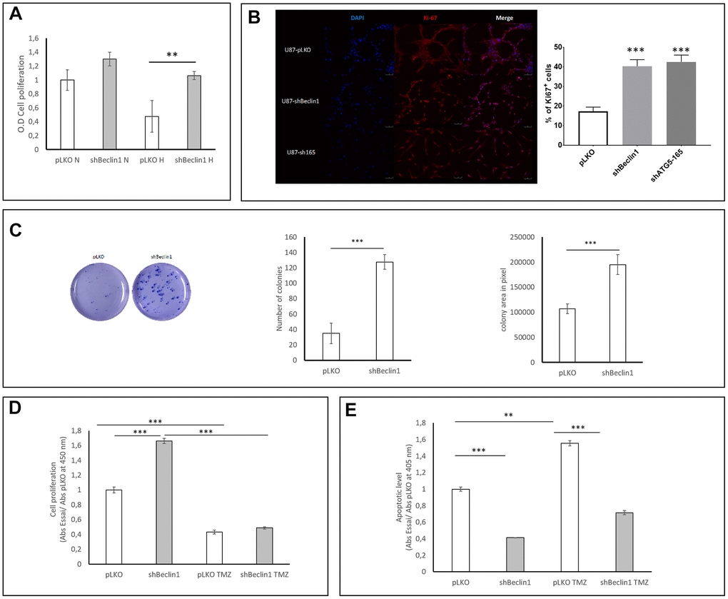
Figure 4.Impact of Beclin1 extinction on glioma cell proliferation and clonogenicity. (A) Cell proliferation in normoxic (N) and hypoxic (H) conditions. Cell proliferation was analyzed using BrdU incorporation (N=3). A slight increase in proliferation rate of U87hBeclin1 cells was observed, as compared with U87pLKO in normoxia; in hypoxic conditions a more significant difference was recorded **p<0.01. (B) Ki67 expression significantly increases in shBeclin1 and shATG5 cells compared to pLKO condition ***p<0,001 (C) Colony forming unit assay with U87shBeclin1. The clonogenic assay was performed as described in material and methods (N=3). There is a significant increase in number (left panel) and size (right panel) of U87shBeclin1 colonies as compared with those obtained with U87pLKO *** p<0.001. (D) Impact of TMZ on cell proliferation. In basal condition, after 144 h of cell culture and 3 h of BrdU incorporation, U87shBeclin1 cells proliferate more than U87pLKO (***p<0.001). TMZ treatment at 1.5 mM induces a very significant decrease in cell proliferation of the two cell lines (*** p<0.001) compared to non-treated cells. There was no difference in proliferative rates between U87pLKO and U87shBeclin1 cells when treated with TMZ (N=3). (E) Impact of TMZ on apoptosis. Apoptotic cell death was evaluated using the Elisa Cell death kit as described in material and methods. After 144 h of culture in basal condition, U87pLKO appears to be the most sensitive cell line to basal apoptosis compared to U87shBeclin1 (***p<0.001). TMZ treatment induces a significant increase in cell apoptosis of U87pLKO cell line (**p<0.01) whereas U87shBeclin1 cells appear to be resistant to TMZ-induced apoptosis (N=3).