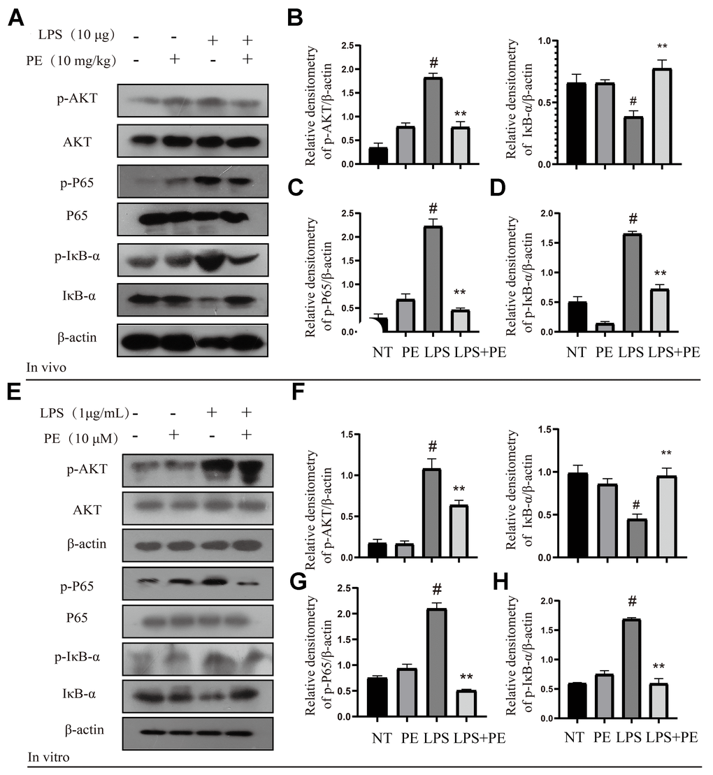
Figure 3.Effect of PE on AKT/NF-κB signal pathway in mastitis models in vivo and in vitro. PE was given orally for 7 days. The fourth pair of milk ducts in mice were injected with LPS for 24 h. The mice were killed by dislocation and fixed on the operating platform after LPS injection 24 h. The midline of abdomen was cut to collect mammary gland. PE was added to the cell culture medium at a concentration of 10 μM. After 1 h, LPS was added to the culture medium at a concentration of 1 μg/mL. The co-stimulation time was 12 h. (NP40) was added to mice mammary gland and mMECs, and then Western blot samples were prepared to obtain protein bands. (A–D) Western blot assay of p-AKT, AKT, p-P65, P65, p-IκB-α and IκB-α in mammary gland. (E–H) Western blot assay of p-AKT, AKT, p-P65, P65, p-IκB-α, IκB-α in mMECs. Each immunoreactive band was digitized and expressed as a ratio of the β-actin level. Values are presented as means ± SEM, three independent repeated experiments were performed. #p<0.01 vs. NT group; **p < 0.01 vs. LPS group.