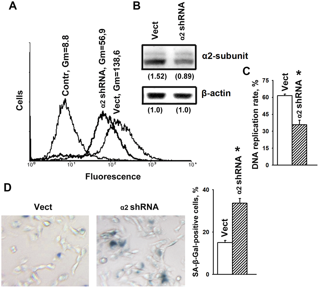
Figure 1.α2β1 deficiency caused senescence of SK-Mel-147 melanoma cells. The cells were transduced with the lentiviral plasmid vector pLKO.1-puro containing α2 shRNA or scramble shRNA (Vect) and selected using puromycin. (A) FACS analysis of α2β1 cell surface expression. Contr: scramble shRNA transduced cells were stained with FITC-conjugated anti-mouse IgG; Vect: scramble shRNA transduced cells were probed with α2β1 mAb and FITC-conjugated anti-mouse IgG; α2 shRNA: α2 shRNA transduced cells were probed with α2β1 mAb and FITC-conjugated anti-mouse IgG; Gm, geometric mean fluorescence intensity. Shown are representative FACS histograms. (B) Western-blotting of the cellular lysate proteins. Cell lysate proteins were run on SDS-PAGE as described in Materials and Methods. The blots were probed with 1:1000 dilution of antibodies to the specified proteins and treated as described in Materials and Methods. Numbers below the bands indicate the α2 band densities normalized against β-actin. Shown are representative blots. (C) α2β1 downregulation reduced the rate of DNA replication of SK-Mel-147 cells. The cells were transduced with scramble shRNA or α2 shRNA and treated as described in Materials and Methods. DNA replication is presented as the ratio (%) Alexa-488-stained cells/Hoechst-stained cells. The results of three independent experiments are shown (M ± SEM); *, ρ < 0.05. (D) SA-β-Gal staining. Cells staining positive for SA-β-Gal showed blue color sedimentation; magnification: × 200. Quantification was performed by counting 200 SA-β-Gal-positive cells in duplicate plates and is presented as a percent of total cells. The results of three independent experiments are shown (M ± SEM); *, ρ <0.05.