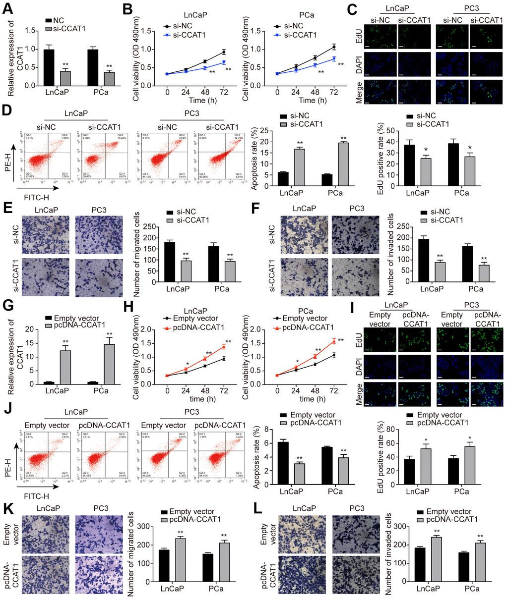
Figure 2.CCAT1 promoted cell proliferation, migration, and invasion in PCa cells. (A) CCAT1 expression in LnCaP and PC3 cell lines. Transfection of si-CCAT1 successfully decreased CCAT1 expression in PCa cells. (B) Cell viability of LnCaP and PC3 cells treated with si-NC or si-CCAT1. CCAT1 inhibition suppressed PCa cells viability. (C) EdU staining in LnCaP and PC3 cells treated with si-NC or si-CCAT1. CCAT1 inhibition suppressed cell proliferation in PCa cells. (D) Cell apoptosis of LnCaP and PC3 cells by flow cytometry detection. PCa cells with lower CCAT1 expression had higher apoptosis rate. (E, F) Cell migration and invasion of LnCaP and PC3 cells in Transwell assays. With si-CCAT1 transfection, cell migration and invasion in PCa cells were both blocked. (G) CCAT1 expression in LnCaP and PC3 cell lines. Transfection of pcDNA-CCAT1 successfully increased CCAT1 expression in PCa cells. (H) Cell viability of LnCaP and PC3 cells treated with empty vector or pcDNA-CCAT1. CCAT1 overexpression enhanced PCa cell viability. (I) EdU staining in LnCaP and PC3 cells treated with empty vector or pcDNA-CCAT1. CCAT1 overexpression promoted cell proliferation in PCa cells. Scale bar: 50 μm. (J) Cell apoptosis of LnCaP and PC3 cells by flow cytometry detection. PCa cells with higher CCAT1 expression level indicated lower apoptosis rate. (K, L) Cell migration and invasion of LnCaP and PC3 cells in Transwell assays. With pcDNA-CCAT1 transfection, cell migration and invasion in PCa cells were both enhanced. *P < 0.05, **P < 0.01, compared with the si-NC group or the empty vector group.