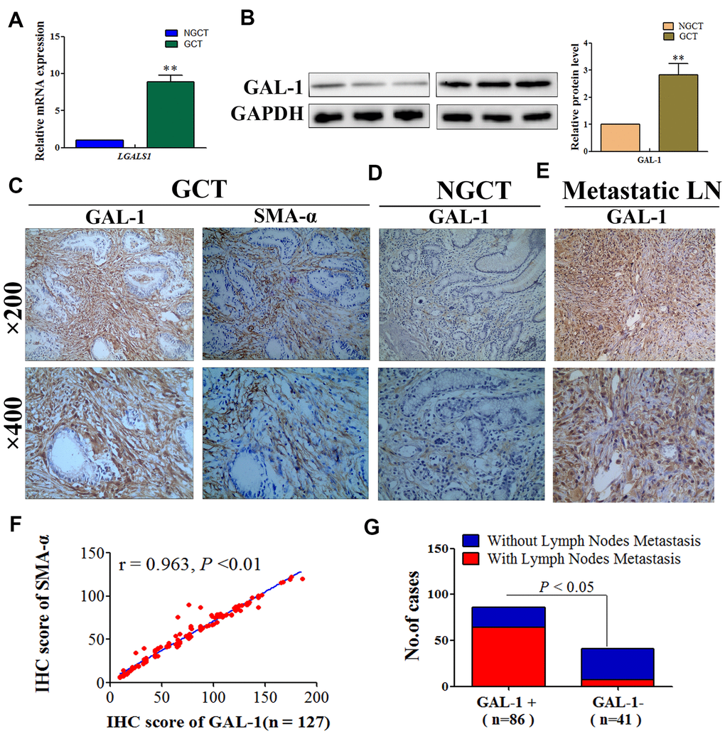
Figure 1.GAL-1/LGALS1 is overexpressed in CAFs and promotes lymph node metastasis in GC tissues. (A) GCT exhibited significantly higher levels of the LGALS1 mRNA than that in NGCT. (B) GAL-1 is overexpressed in GC tissues. (C–E) Representative images of IHC for GAL-1 and SMA-α protein levels in GCT, NGCT, and metastatic LN. (F) The IHC score of GAL-1 correlated positively with the IHC score of SMA-α in GC tissues (r = 0.963; P < 0.01). (G) The lymph node metastasis rate of the GAL-1-positive group was significantly higher than that in the GAL-1-negative group (P < 0.01).