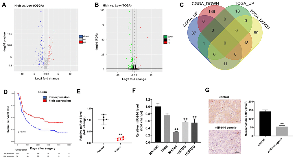
Figure 1.Identification of differentially expressed miRNAs in gliomas from CGGA and TCGA datasets. (A, B) Volcano plots show upregulated (red dots) and downregulated (blue dots) miRNAs in the glioma datasets from (A) CGGA and (B) TCGA databases. (C) Venn diagram shows the numbers of upregulated and downregulated miRNAs or DEMs that are common to both CGGA and TCGA datasets. (D) Kaplan-Meier survival curves show overall survival of glioma patients with low (n=114; blue line) or high (n=76; red line) miR-944 expression in the CGGA dataset. (E) RT-qPCR analysis shows miR-944 expression in glioma tissues and adjacent normal tissues. **P < 0.01 vs. Normal group. (F) RT-qPCR analysis shows miR-944 expression in HA1800 cells and human glioma cell lines, T98G, SHG44, U87MG, and U251MG. **P < 0.01 compared to the HA1800 group. (G) Immunohistochemical (IHC) staining results show CD31 staining of xenograft glioma cell tumor tissues derived from subcutaneous injections of SHG44 cells into nude mice. As shown, positive CD31 staining shows microvessel density in control, agomiR-944- or agomiR-NC-injected xenograft glioma cell tumors at 3 weeks. The tumors were directly injected with 50 nM agomiR-944 or agomiR-NC, twice every week for 3 weeks. **P < 0.01 vs. control group.