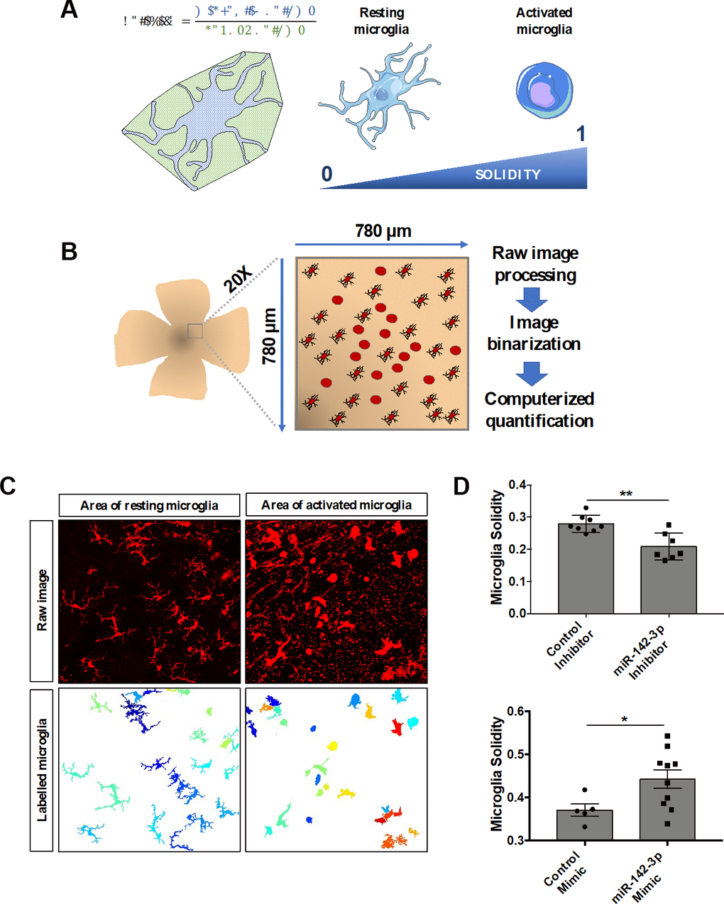
Figure 4.MiR-142-3p influences microglia cell activation state in vivo. (A) Characterization of microglia morphology via cell solidity. The solidity of an object is defined as the ratio between its volume and its convex volume. Resting microglia are highly ramified while activated microglia present an amoeboid shape, with no or small ramifications. Activated microglia are characterized by a higher solidity. (B) CNV lesion area of flat-mounted retinas and surrounding healthy tissue were imaged at the 20X magnification and then processed and quantified. (C) Representative raw images and corresponding labelled images of resting and activated microglia area. (D) Microglia activity measured around the CNV lesion in mice injected with either miR-142-3p inhibitor or mimic and relative controls (n = 5-10 per experimental group). All results are presented as mean +- SEM. Mann Whitney test (* = p ≤ 0.05; ** = p ≤ 0.01).