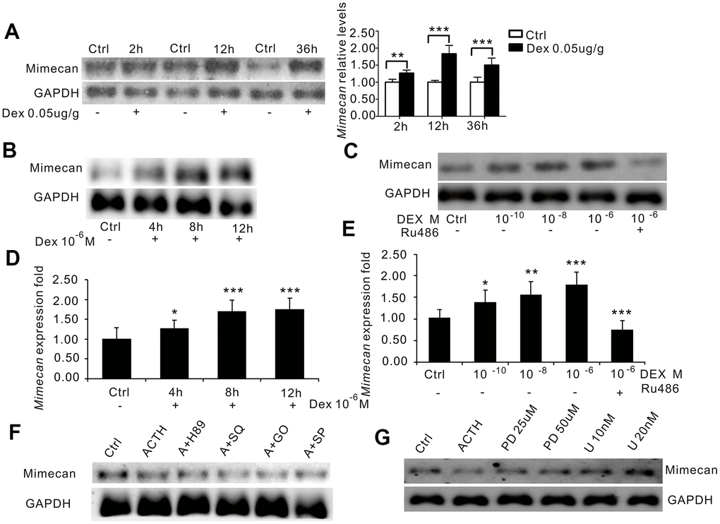
Figure 4.Dexamethasone (DEX) increases mimecan expression in mice and Y-1 cells. (A) Northern blot analysis reflecting the time-dependent increase in mimecan mRNA expression in the adrenal tissues of the C57BL/6 male mice after intramuscular injection of DEX compared with the corresponding control groups that were injected with 0.9% saline (0.05 μg/g; 10 mice per group for each time point). (B) The time-dependent increase in mimecan mRNA in Y-1 cells after DEX treatment (10−6 M), analyzed using Northern blot analysis. Y-1 cells were serum-deprived overnight before adding DEX or DEX + RU486. (C) The dose-dependent increase in mimecan mRNA in Y-1 cells after DEX treatment was abolished by 1 μM RU486, analyzed using Northern blot analysis. (D, E) The mRNA levels in (B) and (C) were determined using quantitative real-time PCR. Relative mimecan mRNA levels were normalized to GAPDH mRNA expression and compared with untreated controls. (F) ACTH-induced suppression of mimecan expression cannot be attributed to PKA, cAMP, PKC, or JNK signaling. (G) The inhibitory effect of ACTH was abolished by the ERK pathway inhibitors PD98059 and U0126, which rescued mimecan expression in a dose-dependent manner. H89: inhibitor of the PKA pathway; SQ: SQ22536, inhibitor of the cAMP pathway; G0: G06983, inhibitor of the PKC pathway; SP: SP600125, inhibitor of the JNK pathway; PD: PD98059, inhibitor of the ERK pathway; U: U0126, inhibitor of the ERK pathway. Y-1 cells were serum-deprived overnight prior to adding inhibitors. Mimecan gene expression in Y-1 cells was analyzed by Northern blot analysis. The relative mimecan mRNA levels were normalized to GAPDH mRNA expression. Data information: *p<0.05, **p<0.01, ***p<0.001 for DEX treatment vs. control, Student’s t-test.