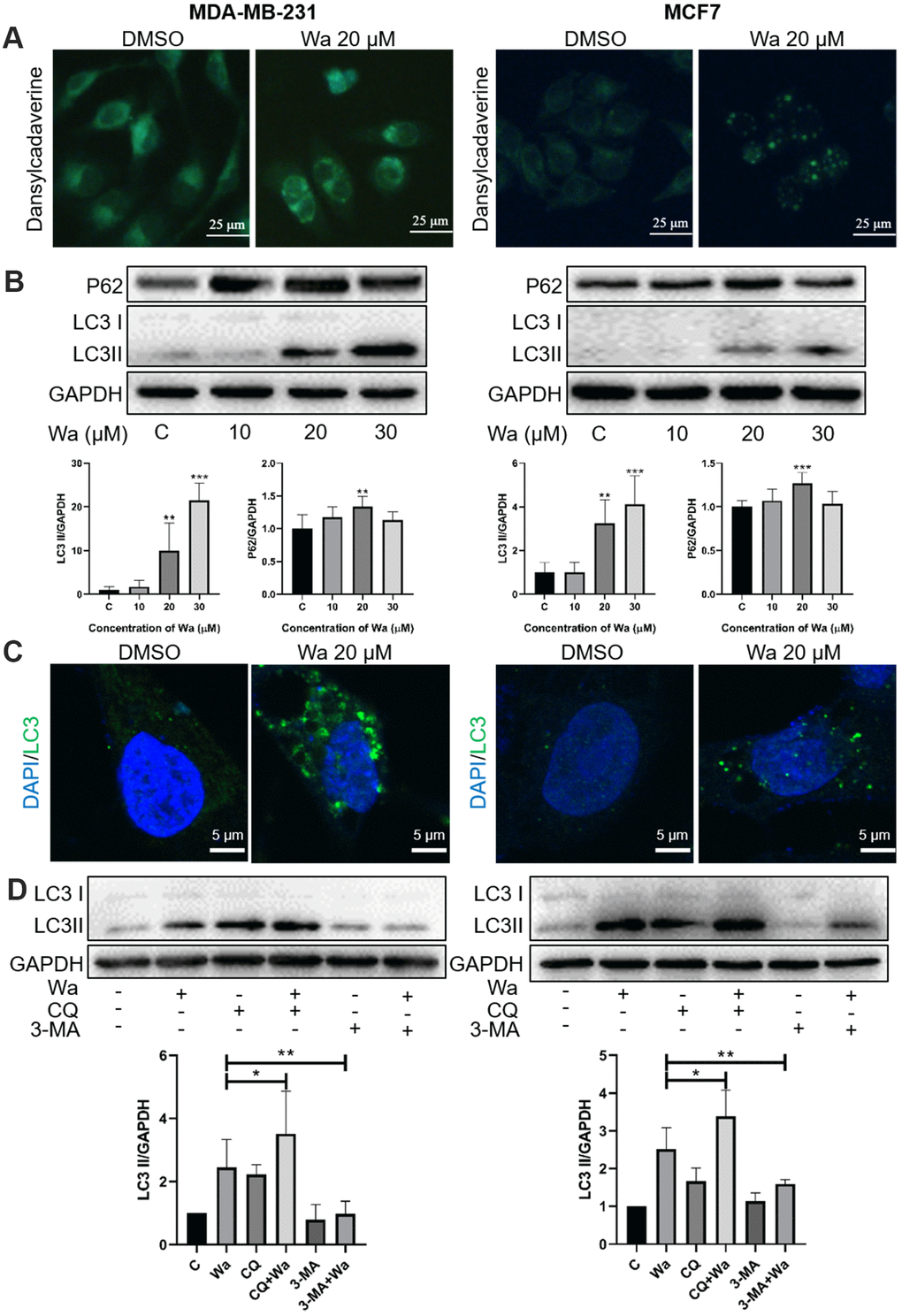
Figure 4.Warangalone induces cell autophagy. (A) MDA-MB-231 and MCF7 cells were treated with 20 μM warangalone for 12 h. Treated and untreated cells were stained with MDC solution and observed by fluorescence microscopy. MDC-labeled autophagic vacuoles emit bright green. (B) MDA-MB-231 and MCF7 cells were treated with the indicated concentrations of warangalone for 12 h. Representative Western blots show the expression of P62 and LC3. GAPDH was used as the loading control. Protein expression of P62 and LC3 II was quantified by densitometry and normalized to GAPDH (ratio of P62 or LC3 II to GAPDH). (C) MDA-MB-231 and MCF7 cells were treated with 20 μM warangalone for 12 h, then immunostained with LC3 antibody and stained with DAPI. Fluorescence was observed with a confocal laser scanning microscope (CLSM). LC3 labeled with Alexa Fluor 488 emits green, and nuclei labeled with DAPI emit blue. (D) MDA-MB-231 and MCF7 cells were pretreated with CQ and 3-MA for 1 h, and then treated with 20 μM of warangalone for 12 h. Representative Western blots show the expression of LC3. GAPDH was used as the loading control. Protein expression of LC3 II was quantified by densitometry and normalized to GAPDH (ratio LC3 II: GAPDH). One-way ANOVA was used for statistical analysis (n ≥ 3). *P < 0.05, **P < 0.01, ***P < 0.001 compared to the respective control group.