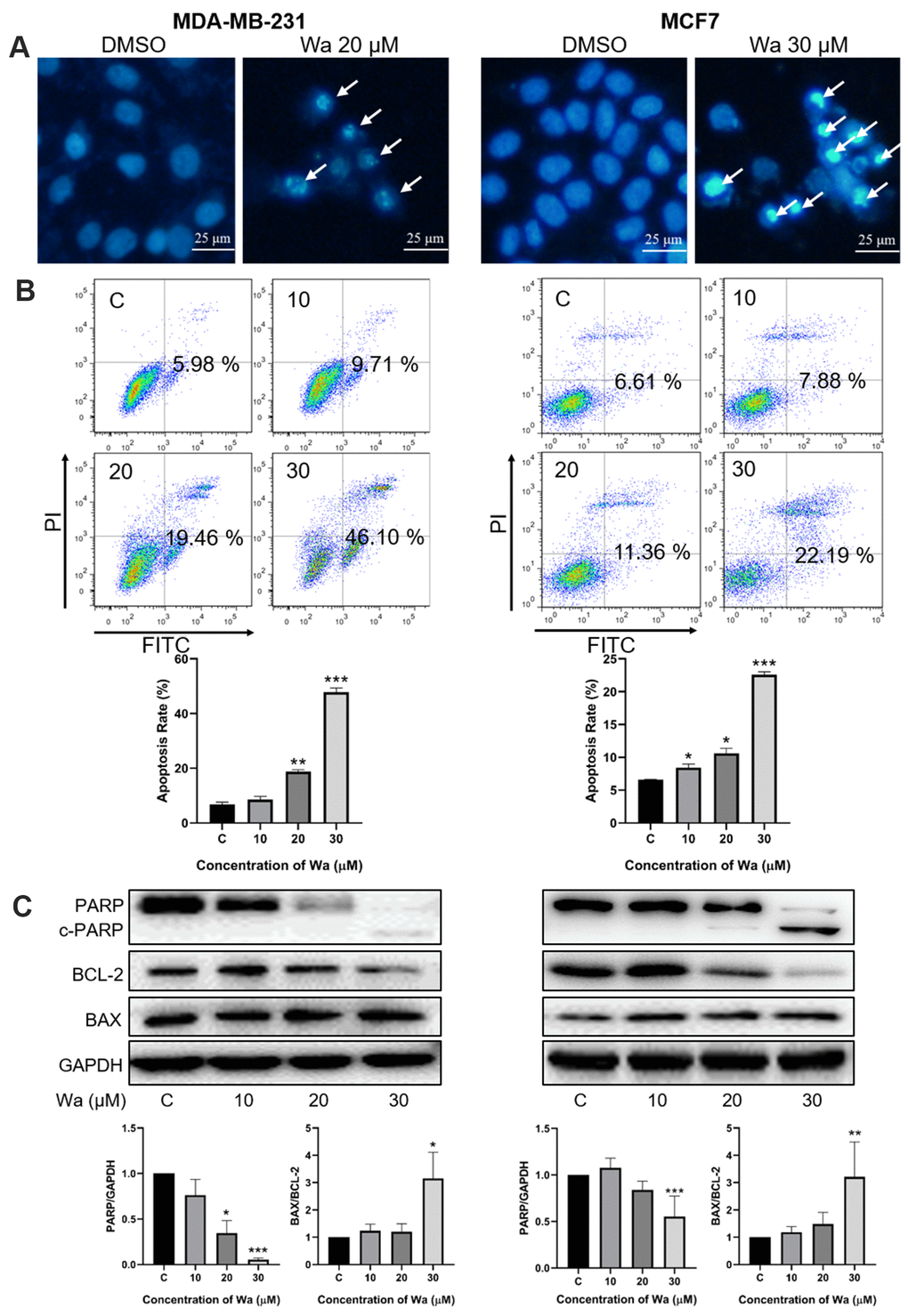
Figure 3.Warangalone induces mitochondrial apoptosis. (A) MDA-MB-231 and MCF7 cells were treated with 20 μM warangalone for 24 h. Treated and untreated cells were stained with Hoechst 33342 solution and the nuclei were observed with fluorescence microscopy. Arrows indicate shrunken and darker nuclei. (B) MDA-MB-231 and MCF7 cells were treated with the indicated concentrations of warangalone for 24 h. The apoptosis rate was detected by Annxin V-FITC/PI assay. (C) MDA-MB-231 and MCF7 cells were treated with the indicated concentrations of warangalone for 24 h. Representative Western blots showed the expression of PARP, BCL-2, and BAX. GAPDH was used as the loading control. Protein expression of PARP was quantified by densitometry and normalized to GAPDH (ratio PARP:GAPDH). Protein expression of BAX was quantified by densitometry and normalized to BCL-2 (ratio BAX:BCL-2). One-way ANOVA was used for statistical analysis (n ≥ 3). *P < 0.05, **P < 0.01, ***P < 0.001 compared to the respective control group.