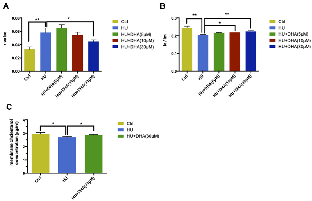
Figure 4.DHA intervention ameliorated membrane aging phenotypes. TMA-DPH and DPP were used as two fluorescent probes to detect membrane mobility in vitro. In the TMA-DPH probing experiment, HU treatment considerably increased the r value compared with the control (P < 0.01). However, co-treatment with different concentrations of DHA gradually decreased the r value. Especially after 30μM DHA treatment, the r value showed a dramatic decline compared with HU treatment (P < 0.05) (A). Similar results were confirmed by the DPP probing test. HU treatment decreased the Ie/Im ratio noticeably (P < 0.01). The ratio gradually increased after co-treatment with the increasing concentrations of DHA. Ie/Im was considerably augmented after treatment with 10μM and 30μM DHA compared with HU treatment (P < 0.05 and P < 0.01) (B). The r value is inversely proportional to membrane fluidity, while the ratio of Ie/Im is directly proportional to membrane mobility. Regarding membrane lipid composition in vitro, the cholesterol level in membrane pellets significantly increased in the 30μM DHA co-treatment group compared with HU treatment group (C). All the data are expressed as mean ± SD from three independent experiments (N = 3). *P < 0.05, **P < 0.01. Student t test and one-way ANOVA were used to determine the statistical significance of the differences.