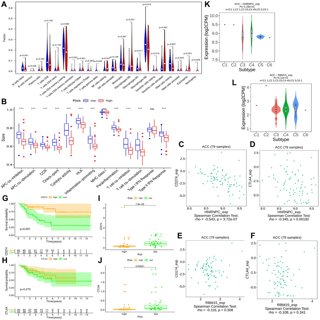
Figure 7.The effect of m6A-related risk signature on ACC immune microenvironment. (A) Comparison of the infiltrating levels of 22 immune cells between different risk groups. (B) Comparison of the activity scores of 13 immune-related pathways between different risk groups. (C–D) The relationships between HNRNPC expression and CD274 (PD-L1), CTLA4 expressions. (E–F) The relationships between RBM15 expression and CD274 (PD-L1), CTLA4 expressions. (G–H) The prognostic differences of ACC patients in TCGA cohort between high- and low-CD274 or CTLA4 expression. (I–J) The expressive difference of CD274 or CTLA4 between high and low m6A-risk group. (K–L) The distributions of m6A risk genes in different PAAD immune subtypes. DC, dendritic cell; APC, antigen-presenting cells; CCR, cytokine-cytokine receptor; IFN, interferon.