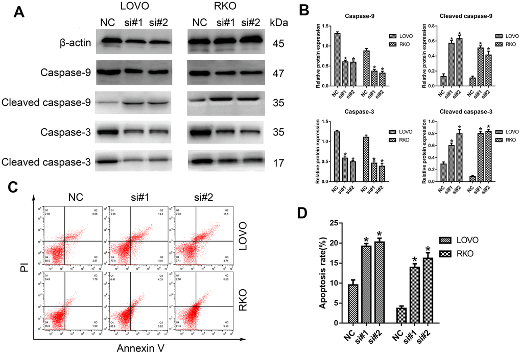
Figure 3.Knockdown of LINC00665 induced apoptosis in CRC cells. (A) Apoptosis-related protein levels were analyzed by Western blotting (n = 6; *P < 0.05 vs NC). (B) Western blotting showed the protein levels in both cell lines (n = 6; *P < 0.05 vs NC). (C, D) The apoptosis rates of LOVO and RKO cells after siRNA treatment were detected by flow cytometry.