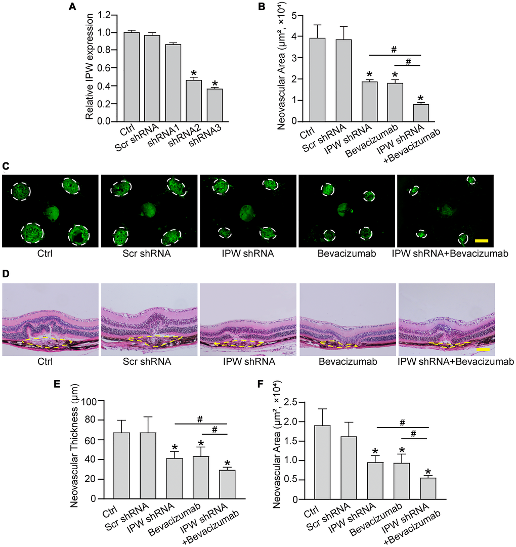
Figure 3.Silencing of lncRNA-IPW inhibits experimental choroidal neovascularization in vivo. (A) C57BL/6 mice received an intravitreal injection of scrambled (Scr) shRNA, IPW shRNA, or left untreated (Ctrl). qRT-PCRs were conducted to detect IPW expression at day 14 after intravitreal injection (n = 6 animals/group; Kruskal-Wallis test). (B–F) C57BL/6 mice received an intravitreal injection of Scr shRNA, IPW shRNA3, bevacizumab, IPW shRNA3 plus bevacizumab, or left untreated (Ctrl). After 14 days, the mice were euthanized and the RPE/choroid complexes were dissected and flat-mounted. The blood vessels were stained by Isolectin-B4 and neovascular area was calculated (B; n = 6 animals/group; Kruskal-Wallis test). (C) The representative images of Isolectin-B4 staining were shown on day 14 after laser photocoagulation. Green staining indicated the CNV lesion. Dashed lines delineate the lesions. Scale bar: 200 μm (C). (D–F) Histological sections of HE stained retinal sections from mice on day 14 after laser photocoagulation. Typical sections of laser-burned eye stained with HE, with the lesion delineated by the dashed line (D). Neovascular degree was estimated by neovascular thickness (E; n = 6 animals/group; Kruskal-Wallis test) and neovascular area (F; n = 6 animals/group; Kruskal-Wallis test). Scale bar: 100 μm. *P < 0.05 versus Ctrl group; #P < 0.05 versus IPW shRNA plus bevacizumab.