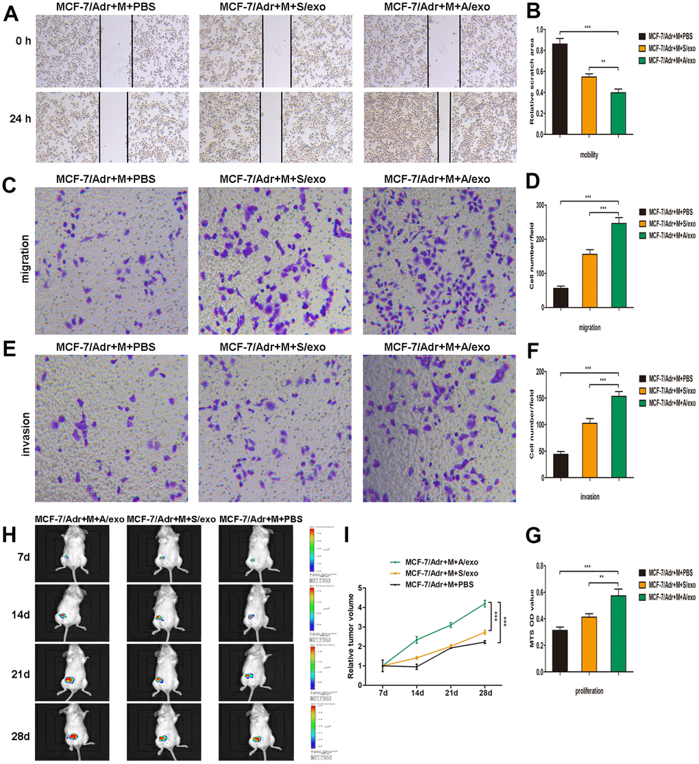
Figure 2.M2 macrophages induced by A/exo promote the mobility, migration, invasion, and proliferation of MCF-7/Adr cells. (A) MCF-7/Adr cells were incubated with culture medium of macrophages (M) treated with PBS, and S/exo or A/exo. Cell mobility of MCF-7/Adr was observed using the wound-healing assay, and representative images of scratch area were shown. (B) Quantitative evaluation of scratch area. (C) Cell migration of MCF-7/Adr was evaluated using the transwell assay, and representative images of migrated cells were shown. (D) Quantitative evaluation of migrated cells. (E) Cell invasion of MCF-7/Adr was determined using the transwell assay, and representative images of invaded cells were shown. (F) Quantitative evaluation of invaded cells. (G) Cell proliferation of MCF-7/Adr was assessed using the MTS viability assay, and quantitative evaluation of OD values were shown. (H) Mice were subcutaneously implanted with a mixture of MCF-7/Adr cells plus macrophages treated with PBS, and S/exo or A/exo. Tumor growth was monitored weekly using a bioluminescence imaging system. (I) Tumor volume was recorded weekly, and tumor growth curve was plotted. Data are shown as mean ± SD, n = 3 independent experiments; * P<0.05, ** P<0.01, and *** P<0.001 compared with controls.