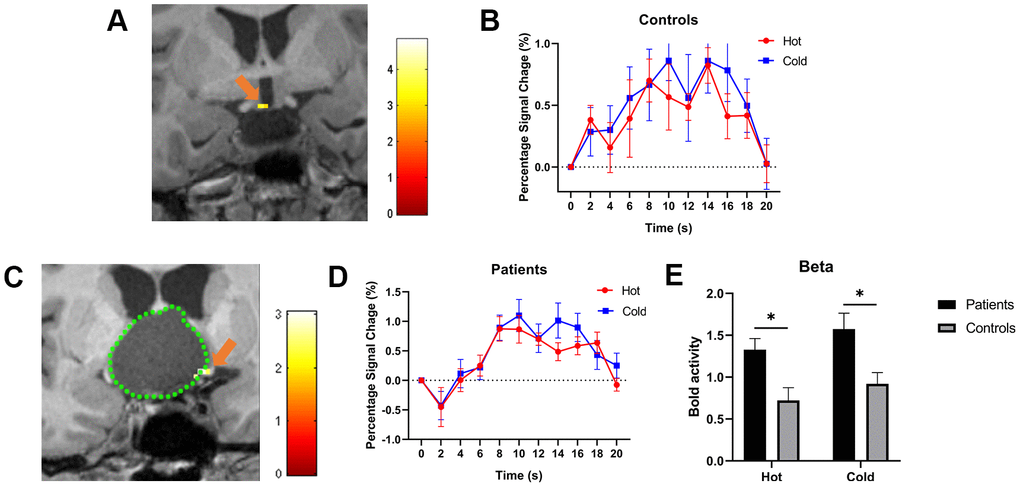
Figure 1.(A, C) show the activated POAH of the hypothalamus in an HV and a CP patient, respectively. The pseudo color indicates the t-value of the activation strength. The orange arrow indicated the activated POAH in the subject. In Figure 1C, the green dotted line was drawn to mark the boundary of the tumor. (B, D) show the BOLD responses to cold and warm stimuli in HVs and patients, respectively. (E) shows the beta values of warm and cold stimuli between patients and HVs. The BOLD activity in patients was significantly higher than that in HVs in response to both cold and warm stimuli. *: P<0.05. POAH: preoptic and anterior hypothalamic region; HV: healthy volunteer; CP: craniopharyngioma; BOLD: blood oxygen level-dependent.
Figure 1 — Predicting the location of the preoptic and anterior hypothalamic region by visualizing the thermoregulatory center on fMRI in craniopharyngioma using cold and warm stimuli | Aging