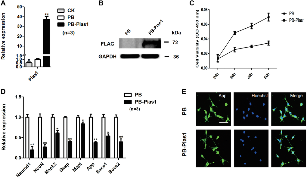
Figure 2.Overexpression of Pias1 regulates AD marker gene expression and cell proliferation in HT-22 cells. (A) qRT-PCR analysis of Pias1 mRNA expression in the hippocampal neuronal HT22 cells transfected with the indicated transgenes. Data represent the mean ± SD of three biological replicates. *p < 0.05, **p < 0.01 versus PB vector. The control check (CK) group represents the baseline expression of Pias1 in HT-22 cells. (B) Western blot analysis of PIAS1 in the hippocampal neuronal HT22 cells with stable Pias1 transgenic expression. (C) Cell proliferation was determined by CCK-8 assays in HT-22 cells transfected with PiggyBac vector (PB) or PB-Pias1. Data represent the mean ± SD. *p < 0.05, **p < 0.01. (D) qRT-PCR analysis of Neurod1, NeuN, Mapk2, Gsap, Mapt, App, Bace1 and Bace2 expression levels in PB and PB-Pias1 hippocampal neuronal HT22 cells. Data are presented as the mean ± SD of three independent experiments. *p < 0.05, **p < 0.01. (E) Immunofluorescence staining of APP in the hippocampal neuronal HT22 cells overexpressing PB-Pias1. Bar: 100 μm.