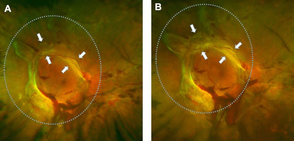
Figure 1.Retinal fibrosis accelerated after IVB treatment. (A) The fundus photograph shows pre-retinal hemorrhage and fibrosis in the macula and optic disc regions of the retina, a circular proliferative membrane presence before IVB treatment. (B) Retinal fibrosis was obviously accelerated post-IVB treatment. The arrows indicate the progression of pre-retinal membrane fibrosis compared to pretreatment conditions.