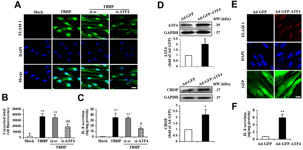
Figure 3.ATF4 is an important mediator of ELAM-1 expression and IL-8 secretion in HTMCs. (A–C) HTMCs were exposed to media in the absence (Mock) or presence of 50 μM of TBHP for 12 h. Alternatively, cells were transfected with si-ATF4 or si-sc for 48 h, followed by incubation with 50 μM of TBHP for additional 12 h. (A) Representative images of subcellular expression of ELAM-1 as examined by indirect immunofluorescence (green, ELAM-1. blue, DAPI. scale bar, 40 μm). (B) Levels of cellular immunofluorescence of ELAM-1 were quantified and expressed as corrected total cell fluorescence (mean ± SEM, n = 3). (C) IL-8 secretion was assayed in culture supernatant of HTMCs using an IL-8 ELISA kit. Values were normalized for total protein at the respective treatment (mean ± SEM, n = 3). **P <0.01 vs. Mock, #P <0.05 and ##P <0.01 vs. TBHP+si-sc. TBHP+si-sc, exposure of cells transfected with si-sc to TBHP. TBHP+si-ATF4, exposure of cells transfected with si-ATF4 to TBHP. (D–F) A recombinant adenovirus coding ATF4 (Ad-GFP-ATF4) was constructed to express GFP protein as a marker for the identification of infected cells. Cultured HTMCs were infected with Ad-GFP-ATF4 or empty vector (Ad-GFP) for 72 h. (D) Western blot analysis for ATF4 and CHOP. Intensities of protein expression were quantified, normalized against the level of GAPDH and expressed as relative changes to Ad-GFP (mean ± SEM, n = 3). (E) Subcellular expression of ELAM-1 was detected by immunofluorescent staining (red, ELAM-1. blue, DAPI. green, GFP. scale bar, 20 μm). (F) IL-8 secretion was assayed using ELISA. Values were normalized for total protein at the respective treatment (mean ± SEM, n = 3). *P <0.05, **P <0.01 vs. Ad-GFP.