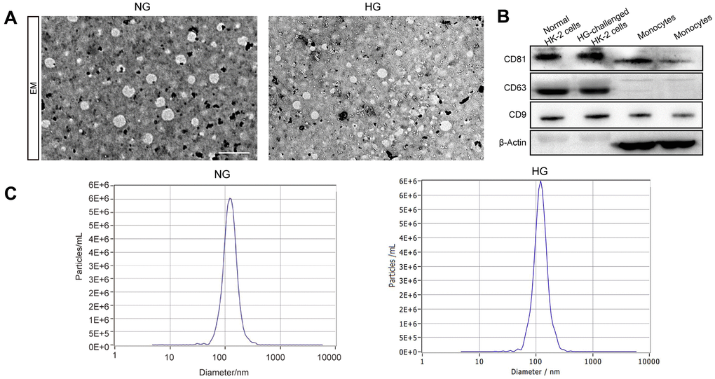
Figure 1.The identification of exosomes. Representative TEM image of exosomes derived from two groups (A). Western blot of the exosomal markers CD63, CD9, and CD81 and non-exosomal protein markers β-Actin in exosomes and monocytes (B). The size of the exosomes (nm) enriched from the culture supernatant of two groups was examined through NTA using a NanoSight NS300 instrument (NanoSight Ltd., Amesbury, UK) (C).