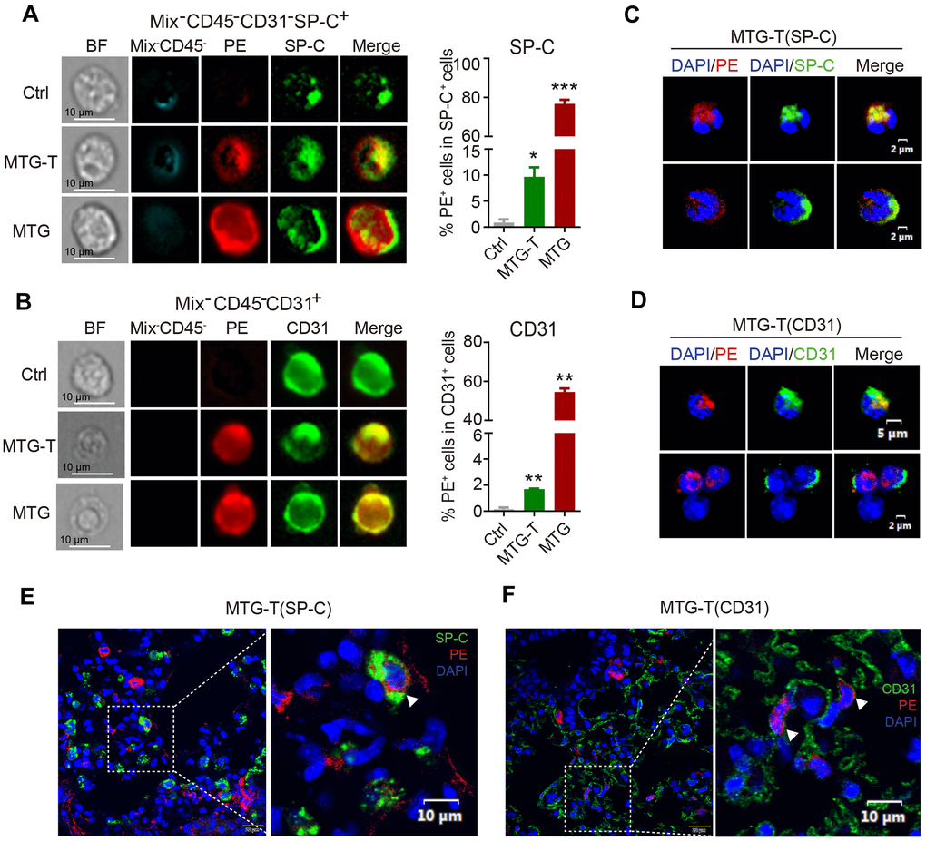
Figure 3.Co-localization of donor-derived cells and recipient lung tissue cells were observed. (A) The representative images by image flow cytometry results of donor derived PE red fluorescence and the recipient lung epithelial cell marker SP-C and (B) endothelial cell marker CD31. The right side showed the quantitative analysis of SP-C with PE, CD31 with PE with exclusion of the blood cells (Blood Mix antibodies and CD45). (C, D) Representative images of single-cell confocal of PE red fluorescence with lung epithelial cell SP-C and endothelial cell CD31 in sorted single PE positive cells of MTG-T group. (E, F) Representative images of confocal co-localizaiton of PE red fluorescence with lung epithelial cell marker SP-C, endothelial cell marker CD31 in situ lung section of MTG-T group, DAPI was stained to characterize the nucleus.