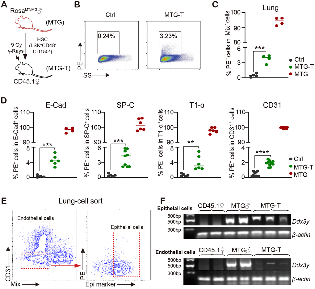
Figure 2.Male donor-derived PE red fluorescence cells and Y chromosome were detected in female recipient lung tissue cells. (A) HSC from male RosamT/mG mice were sorted and transplanted into lethally irradiated female CD45.1 mice. (B) Flow cytometry chart of PE (tdTomato) fluorescence in lung tissue cells with exclusion of blood cells including lineage (CD3, CD8, B220, Gr-1, TER119), macrophage (CD11b, F4/80), megakaryocyte (CD41/CD61) and CD45 (marked as Mix). (C) Statistical analysis of PE positive percentage in lung tissue cells. (D) PE percentage in lung epithelial cells (E-Cadherin, SP-C and T1-α) and endothelial cells (CD31) (after exclusion of blood cells). (E) For Ddx3y detection, lung epithelial cells (Epithelial marker+/blood Mix marker-/CD31-) and endothelial cells (blood Mix marker-/CD31+) were sorted. (F) Representative images of Ddx3y expression by amplification with PCR and detection by nucleic acid electrophoresis. N≥4 in (C, D, E, F). **:p<0.01; ***: p<0.001; ****: p<0.0001.