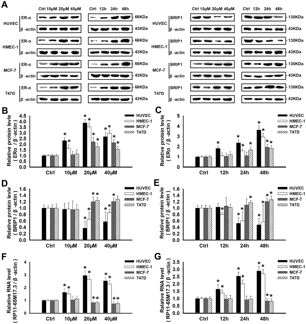
Figure 3.Calycosin regulated cell proliferation by activating or repressing the RP11-65M17.3-ERα loop. (A–E) HUVECs, HMEC-1 cells, MCF-7 cells and T47D cells were treated with calycosin (10, 20 and 40 μM) for 12, 24, or 48 h. Western blotting was used to determine the protein levels of ERα and BRIP1, and β-actin served as the internal control. (F, G) The levels of RP11-65M17.3 were determined by qRT-PCR and normalized to those of β-actin.
(H–M) The effects of calycosin on the RP11-65M17.3-ERα loop were demonstrated by pretreating cells with RP11-65M17.3 shRNA (RI) or MPP. The protein and mRNA expression levels of BRIP1, ERα and RP11-65M17.3 were determined using Western blotting and qRT-PCR. The protein and transcript levels were normalized to the β-actin levels. Representative data from three independent experiments are shown. *p < 0.05 vs. control (0 μM or 0 h); #p < 0.05 vs. 20 μM calycosin alone; $p < 0.05 vs. 10 nM E2 alone.