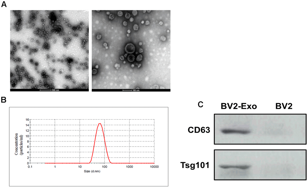
Figure 2.Identification of M2-phenotype microglia-derived exosomes (BV2-Exo). (A) Morphology of BV2-Exo, as determined by transmission electron microscopy. Scale bar = 500 nm (left panel) and 100 nm (right panel). (B) Size distribution of exosomes, as determined by nanoparticle tracking analysis. (C) Expression of exosome markers CD63 and Tsg101 in BV2-Exo and BV2 cells. Data are presented as mean±SD. *, p<0.05. At least three replicates were available for statistical analysis in each treatment.