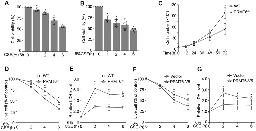
Figure 7.PRMT6 overexpressed epithelia are protected from CSE driven cell death. (A, B) BEAS-2B cells were treated with various concentrations of CSE at indicated time points. Cell viability was measured with MTT assay. (C) 5×10⁴ cells of WT or PRMT6-/- cell were cultured for a variety of time points, then stained by trypan blue and counted by TC10 automatic cell counter. (D) The trypan blue stained cells were counted by TC10 automatic cell counter. The percentage of live cells relative to control group after CSE treatment in WT and PRMT6-/- groups were presented. (E) LDH assay was used to determine cell death induced by CSE treatment in WT and PRMT6-/- groups. Relative LDH activity indicated the epithelial cell death rate were plotted. (F) 3 μg of pcDNA3.1D-PRMT6-V5 plasmid and control plasmid were transfected into BEAS-2B cells via electroporation. After 48h, the vector and PRMT6 overexpressed BEAS-2B cells were treated with CSE at various time points. The percentage of live cells were counted and compared with control. (G) Relative LDH activity under CSE treatment in vector and PRMT6-V5 overexpressed group were determined with LDH assay. Results were shown as mean ± SD and representative of n≥3 experiments. Statistics were measured by 1-way and 2-way ANOVA or Student t test, *p < .05 indicated the statistical significance.