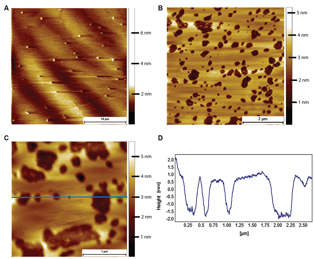
Figure 7.AFM tapping mode images of mouse brain tissue derived supported lipid bilayers in PBS exposed to 8 μM of human Aβ for 2 hours. (A) Large scale image showing a sparse amount of phase separation prior to Aβ adsorption. (B) Mouse brain derived lipid bilayer after exposure for 2 hours to 8 μM of human Aβ. Pits appear in the membrane, with some structure visible within the pits. (C) A close up of the pits showing that they do not break through the entire bilayer, i.e. they are not holes. (D) Line profile corresponding to the horizontal blue line in (C). The pits are between 2 and 2.5 nm deep, too shallow to be holes in the bilayer, which is ~3.1 nm thick.