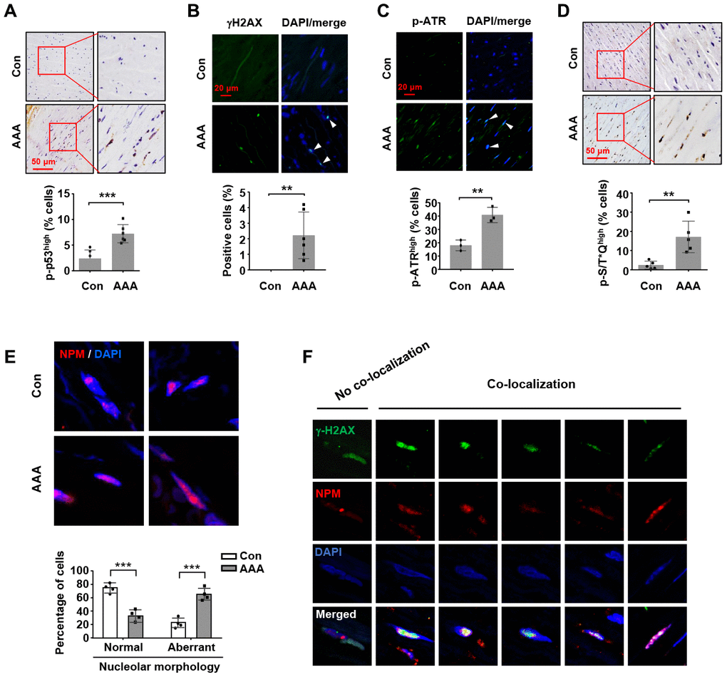
Figure 6.Increases in nucleolar stress response, p53 phosphorylation and DNA damage response in human AAA tissues. (A) Immunohistochemical staining and semi-quantitative data showing the increased level of p53 phosphorylation in the medial layer of normal (Con) and AAA aortas (n = 6). (B) Immunofluorescence results showing the increased number of γH2AX foci (arrowheads) in AAA tissues (n = 6). (C) Immunofluorescence results showing the increased number of cells exhibiting a high level of ATR phosphorylation (arrowheads) in AAA tissues (n = 3). (D) Immunohistochemical staining and semi-quantitative data showing the increased level of phospho-S/T*Q motif of ATM/ATR substrates in AAA tissues (n = 5). (E) Immunofluorescence images of NPM staining showing medial cells with normal nucleoli morphology (examples from control aortas) and those with aberrant nucleoli morphology (highly diffused NPM fluorescence signal) (examples from AAA tissues). The quantitative data below showed the proportions of cells with normal and aberrant nucleoli morphology in control and AAA aortas (n = 4). (F) Example images of AAA medial cells positive for γH2AX foci showing non-co-localization and co-localization of γH2AX with NPM. Dot blot-combined bar graphs represented mean ± S.D. ** P < 0.01, *** P < 0.001 versus control, unpaired t-test.