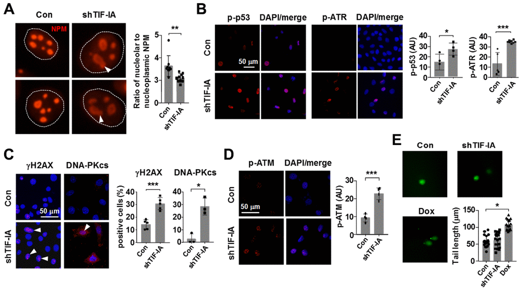
Figure 5.Nucleolar stress elicited a DNA damage response in vascular SMCs (MOVAS). (A) Immunofluorescence staining for nucleophosmin (NPM) showing that TIF-IA silencing triggered a nucleolar stress response as evidenced by the appearance of nucleolar caps (arrowheads) and redistribution of NPM from nucleoli to the nucleoplasmic space. Single nuclei were outlined by the dashed line. The graph on the right showed changes in the ratio of nucleolar to nucleoplasmic NPM fluorescence intensity (n = 9 - 10). (B) Immunofluorescence images and semi-quantitative mean intensity data expressed in arbitrary units (AU) showing the increased phosphorylations of p53 and ATR in TIF-IA-silenced cells (n = 3 - 8). Nuclei were counterstained with DAPI (blue). (C) Immunofluorescence images and quantitative data showing accumulation of γH2AX and DNA-PKcs foci (arrowheads) in the nuclei of TIF-IA-silenced cells (n = 3 - 5). (D) Immunofluorescence and the mean intensity data showing the increased phosphorylation of ATM in TIF-IA-silenced cells (n = 4). (E) Alkaline comet assay results showing that TIF-IA knockdown did not cause massive DNA breaks (n = 17 cells measured). Doxorubicin (Dox, 1 μM) was used as a positive control. Data were mean ± S.D. * P < 0.05, ** P < 0.01, *** P < 0.001 versus control (Con), unpaired t-test or one-way ANOVA as appropriate.