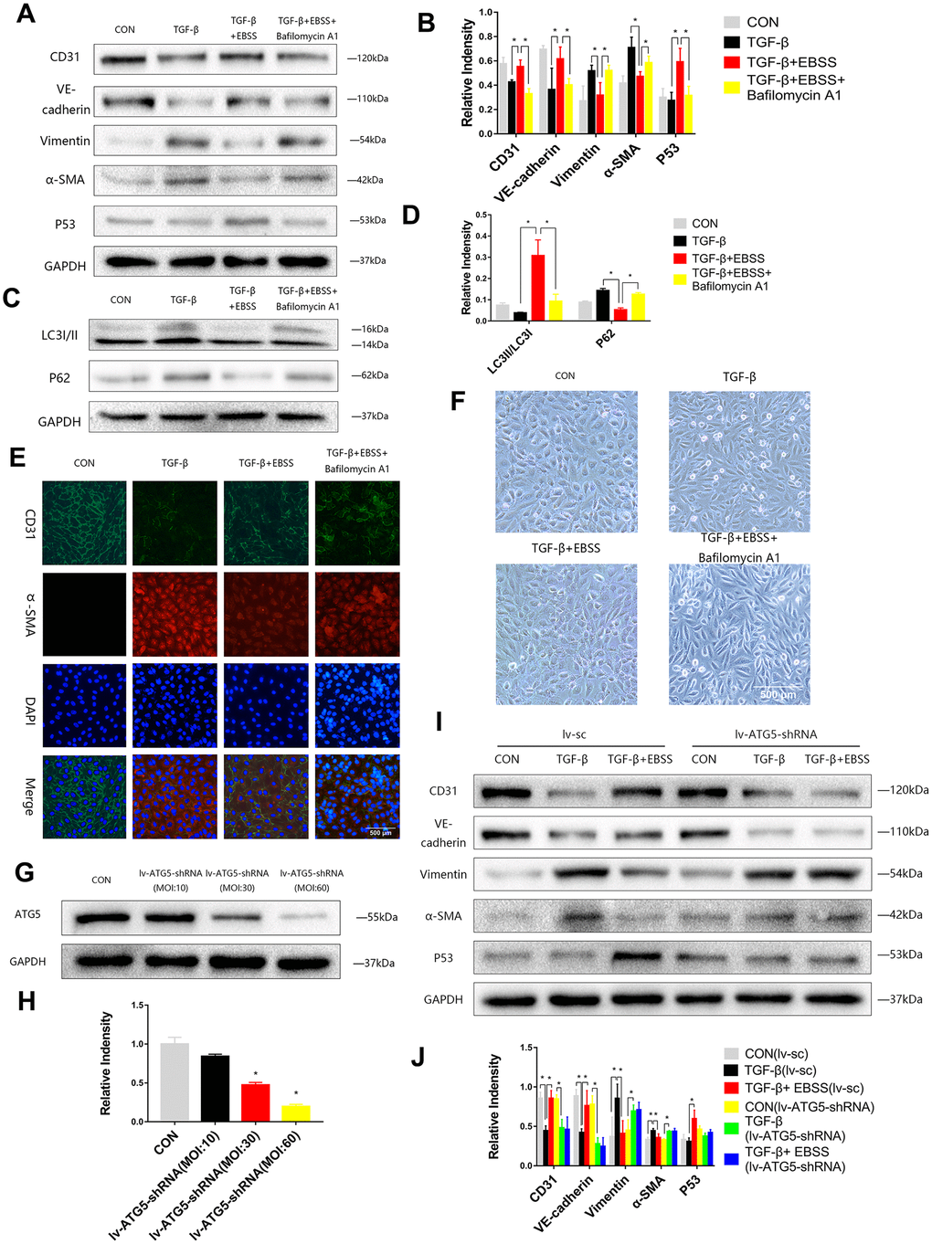
Figure 2.Autophagy modulated MEndT. (A–D) Cells were subjected to TGF-β (20 ng/ml) for 24 h and then treated with EBSS for 4 h, with pretreatment with bafilomycin A1 for 2 h or not. CD31, VE-cadherin, Vimentin, α-SMA, LC3-II/LC3-I, p62 and p53 levels were analyzed by immunoblot. (E) MEndT was assessed by representative images (Scale bars= 500 μm) of immunofluorescence after staining of CD31 and α-SMA. (F) Representative microphotographs (Scale bars= 500 μm) of HUVEC morphological changes. (G, H) Cells were infected with GFP-labeled lv-ATG5 for 24 h, and infection was assessed by the expression of ATG5. (I, J) Cells were treated with TGF-β and EBSS as described above. CD31, VE-cadherin, Vimentin, α-SMA and p53 levels were analyzed by immunoblot. Bar graphs represent data from three independent experiments, and data represent the means±SEM. Unpaired T test (*P<0.05) was used to compare the significances between two groups.