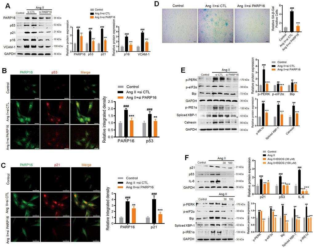
Figure 3.Knocking down or inhibition of PARP16 mitigates Ang II-induced RAECs senescence and endoplasmic reticulum (ER) stress. (A–E) PARP16 knockdown reversed Ang II-induced RAECs senescence and endoplasmic reticulum stress. Senescence-associated markers (VCAM-1, p16, p21, and p53) were assayed by Western blot for RAEC cells transfected with control (si CTL) or PARP16 siRNA before and after Ang II (2 μM) treatment for 48 h (A); Immunofluorescence double staining of PARP16 and p53 (B), and Immunofluorescence double staining of PARP16 and p21 (C); SA-β-Gal staining for RAEC cells (D); ER-associated markers (Bip, p-PERK, p-eIF2α, p-IRE1α, spliced XBP-1 and Calnexin) were assayed by Western blot for RAEC cells transfected with control or PARP16 siRNA before and after Ang II treatment (E). (F) PARP16 inhibitor EGCG reversed Ang II-induced RAECs senescence and endoplasmic reticulum stress. p21, p53, IL-6, Bip, p-PERK, spliced XBP-1, p-IRE1α and p-eIF2α were assayed by Western blot for Ang II-induced RAEC cells with or without PARP16 inhibitor (EGCG) at different concentration. GAPDH serves as internal control. All data were shown as mean ± S.D of at least 4 independent experiments. ##p < 0.01, ###p < 0.001 vs. control; *p < 0.05, **p < 0.01, ***p < 0.001, vs. Ang II+si CTL or Ang II treated cells.